
Imaging method for medical diagnostics and device operating according to this method

Inactive Publication Date: 2009-01-01
SIEMENS HEALTHCARE GMBH
View PDF23 Cites 2 Cited by
  • Summary
  • Abstract
  • Description
  • Claims
  • Application Information

AI Technical Summary

Benefits of technology

[0006]An object of the present invention is to provide an imaging method for medical diagnostics that makes the interpretation of image data acquired with endoscopic methods and non-endoscopic methods easier for the user. A further object of the invention is to provide a device operating according to such a method.
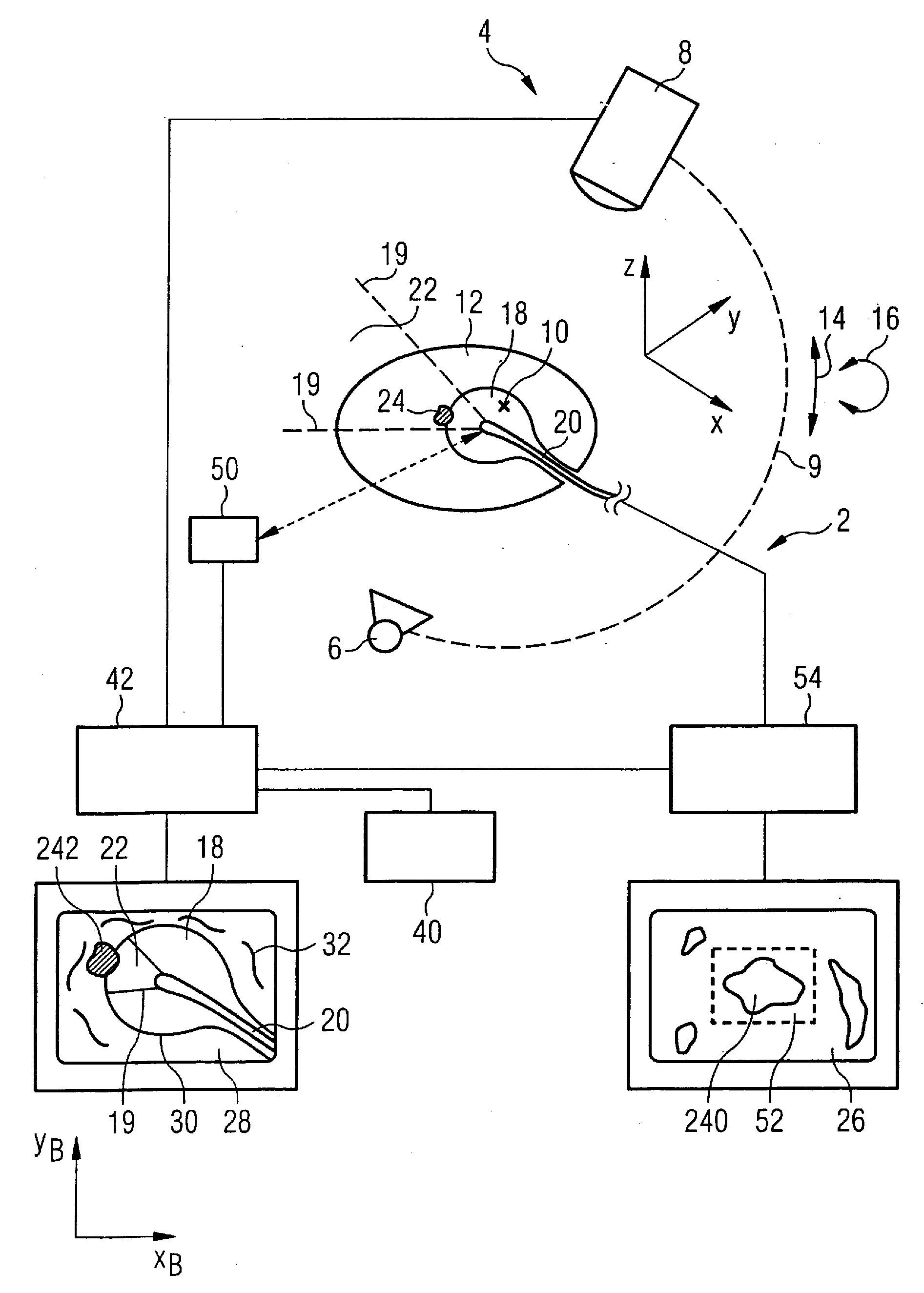
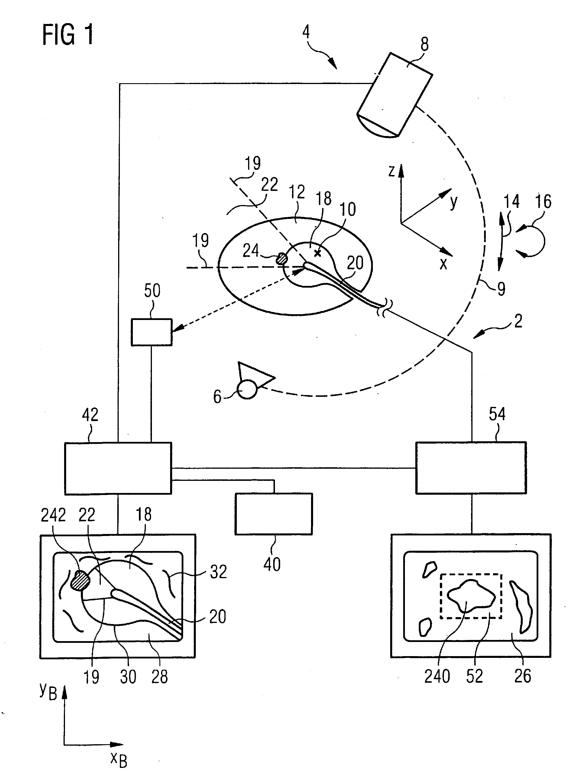
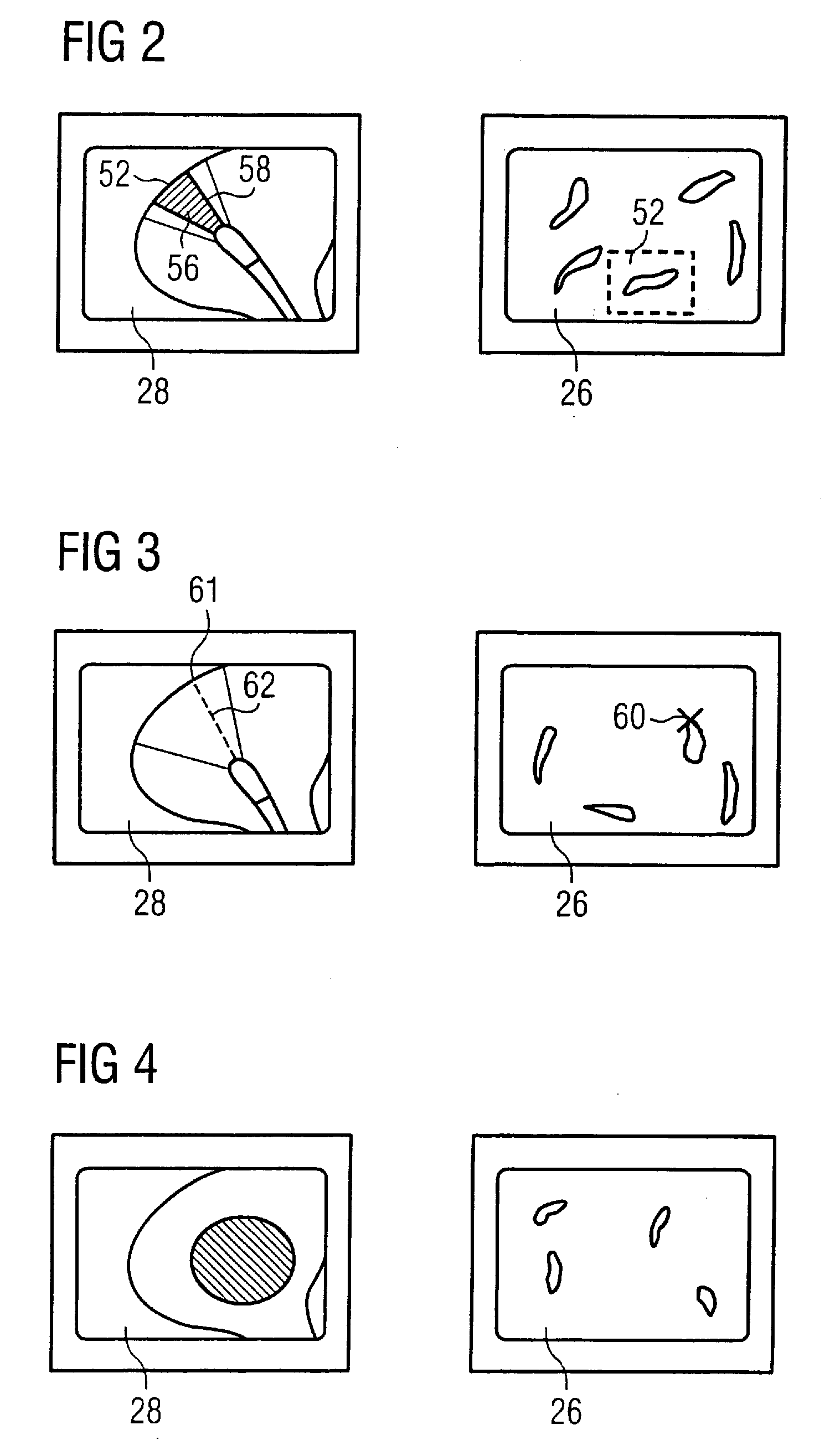
[0012]The association of a structure rendered in the endoscopy image with a structure rendered in the image is made easier for the observer if the rendered image field is bounded by optically impermeable structures recognizable in the image.

Problems solved by technology

Each image, however, offers only two-dimensional information of the state of a section of the inner surface of the cavity and no information from deeper slices or in other viewing directions.
The linking of individual or multiples endoscopic images with one or more x-ray images of the same body require on the part of the observer not only a precise knowledge of the anatomy but also a developed three-dimensional spatial sense that must first be learned and frequently leads to misinterpretations.
Moreover, pathological structures often deviate in a complex manner from the standard, such that a linking of endoscopy images and x-ray images is made more difficult.
One of the causes for these difficulties is that the methods normally used in addition to endoscopy (for example magnetic resonance methods or ultrasound methods in addition to the aforementioned x-ray methods) provide slice images of the examination subject, while in an endoscopy image only a boundary surface between an optically permeable medium and an optically impermeable medium is presented on a two-dimensional image plane.

Method used


Image

  • Imaging method for medical diagnostics and device operating according to this method
  • Imaging method for medical diagnostics and device operating according to this method
  • Imaging method for medical diagnostics and device operating according to this method

Examples


Embodiment Construction

[0020]As shown in FIG. 1, a device according to the invention has an endoscopy apparatus 2 as well as an image generation system 4 operated according to a non-endoscopic method, in the example a C-arm x-ray apparatus with an x-ray source 6 and an x-ray receiver 8 that are arranged on a C-arm 9 (shown in dashes in the Figure). The C-arm 9 can be pivoted around an isocenter 10 such that a two-dimensional image (slice image) can be generated from a body region of a patient 12. This pivot movement around an axis perpendicular to the plane spanned by the C-arm 9 (the plane of the drawing in FIG. 1) is illustrated by a double arrow 14. Moreover, if the C-arm 9 can be pivoted around an axis that lies in the plane spanned by it and is illustrated by the double arrow 16, it is possible to also generate a 3D image data set from the body region of the patient 12.

[0021]An endoscope 20 with which it is possible to optically observe a section of the internal surface of a wall 30 of a cavity 18 is...


PUM


Abstract

In an imaging method for medical diagnostics and a device operating according to this method, during an endoscopic examination of a body region of a patient with an endoscope, an image is generated with a non-endoscopic imaging method and the image field of the endoscope is determined and rendered in the image (28) with accurate position and orientation.

Description

BACKGROUND OF THE INVENTION[0001]1. Field of the Invention[0002]The invention concerns an imaging method for medical diagnostics as well as an imaging device operating according to this method.[0003]2. Description of the Prior Art[0004]In medical diagnostics, in a series of application cases different imaging methods are used simultaneously or successively in order to facilitate the diagnosis or to avoid misdiagnoses. For example, in urology it is known to implement both an endoscopic examination and an x-ray examination. In an endoscopic method, high-resolution, color, optical images are generated in real time in the viewing direction of the endoscope. Each image, however, offers only two-dimensional information of the state of a section of the inner surface of the cavity and no information from deeper slices or in other viewing directions. In an x-ray method, information is also acquired from regions that are not visible in an endoscopy image. The linking of individual or multiple...

Claims


Application Information

Patent Timeline
IPC IPC(8): A61B1/04
CPCA61B5/06A61B6/12A61B2019/5289A61B2019/5238A61B19/52A61B90/36A61B2090/364A61B2090/376A61B5/064A61B5/065
Inventor FEHRE, JENSKUTH, RAINER
Owner SIEMENS HEALTHCARE GMBH
Learn More