
Method of genetic screening and analysis

a genetic screening and analysis technology, applied in the field of genetic screening and analysis, can solve the problems of increased miscarriage risk, infection or leakage, and increased risks for both women and fetuses, and achieve the effect of increasing the accuracy of prediction

Inactive Publication Date: 2009-01-29
GENECARE MEDICAL GENETICS CENT
View PDF9 Cites 4 Cited by
  • Summary
  • Abstract
  • Description
  • Claims
  • Application Information

AI Technical Summary

Benefits of technology

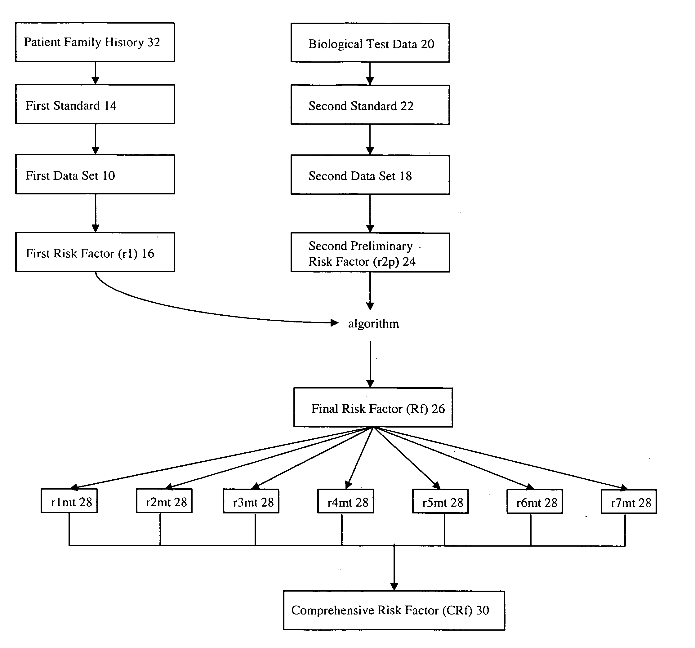
[0007]The present invention is directed to a method for genetic screening and analysis. The first aspect of the present invention is directed to a method of prenatal screening. The method comprises the steps of providing a first data set 10. The first data set 10 comprises patient data 12 of a patient. The patient data 12 is compared to a first standard 14 that provides a first risk factor (r1) 16 for genetic defects. The method further comprises providing a second data set 18. The second data set 18 comprises biological test data 20 from the patient. The biological test data 20 from the patient is compared to a second standard 22 that provides a second preliminary risk factor (r2p) 24 for genetic defects. The method further provides a final risk factor (Rf) 26 based upon an integration of r116 and r2p 24. Rf 26 provides increased accuracy prediction for genetic defects in a fetus of the patient.

Problems solved by technology

Due to the invasive nature of amniocentesis testing, there are potential risks involved including the introduction of pathogens into the amniotic sac from the needle, or the puncture wound to the amniotic sac not healing properly, leading to infections or leakage.
Genetic amniocentesis is generally restricted to being performed no later than the second trimester, and is not performed early in pregnancies because there is not an ample source of amniotic fluid when the fetus is young.
A CVS procedure can be preformed as early as 10-12 weeks, however it has been found to result in increased risks of miscarriages.
Both amniocentesis and CVS are invasive procedures involving risks to both the women and the fetus.
Fetal ultrasound exams in the first or second trimester can screen for anomalies or soft markers associated with certain genetic disorders, but such exams are costly and yield lower detections and higher false positive test results.
One of the drawbacks of current non-invasive methods is that the screening is specific to three chromosome disorders.

Method used


Image

  • Method of genetic screening and analysis
  • Method of genetic screening and analysis
  • Method of genetic screening and analysis

Examples


examples

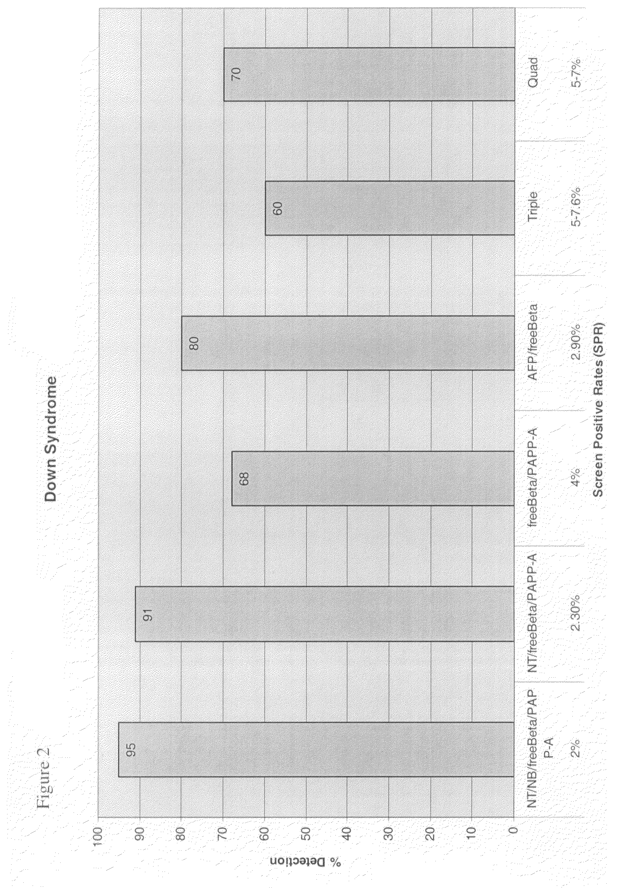
[0022]The following Table is a comparison of prenatal screening programs, including detection rates and screen positive rates for determining the weight of each risk factor. More weight is given for the more effective risk factors. Combining ultrasound measurements for Nuchal Translucency, dried blood freeBeta, and PAPP-A provide higher detection than older methods of screening for Down syndrome and Trisomy 18. Nasal bone or Tricuspid Flow can be added to increase detection by 4-5%.

First Trimester Detection Rates & Screen Positive RatesEarlyResearch / ExperimentalScreen ®Non-FMFWithNT / hCG / PAPP-IntegratedEarly Screen ®NasalA (No USAandFMF / NT / freeBeta / PAPP-ABoneFreeBeta / PPAP-ARef.Sequential*Down Syndrome (DS)91% at 2.3%95% at68% at 4.5%82% at 5-7%Varies*2%(82-86%)Trisomy 1897% at 0.4%98% at>90% at 0.4%No USA Ref.No USA0.4%Ref.Trisomy 1397% at 0.1%98% at90% at 0.1%NoNo0.1%Twins80% at 7.2%80% at50%No USA Ref.No7.2%Heart Defeats / Anomalies40% / Yes40% / No / YesNoNo USAYesRef.Diabetics / Multiples / ...


PUM

PropertyMeasurementUnit
Lengthaaaaaaaaaa
Timeaaaaaaaaaa
Timeaaaaaaaaaa

Abstract

A method of genetic screening and analysis, particularly for prenatal genetic screening to determine a risk estimate or probability of genetic disorders for a specific pregnancy integrating a patient's family history data (r1) with a patient's biological test data (r2p) to generate a final prenatal risk factor (Rf). A comprehensive risk factor (CRf) is generated by running a multiplicity of tests on the Rf for screening various genetic disorders.

Description

TECHNICAL FIELD[0001]The present invention relates to methods of genetic screening and analysis. More particularly, the present invention relates to a method of prenatal screening that generates a final prenatal risk factor (Rf) for genetic defects by integrating a patient's family history data (r1) with a patient's biological test data (r2p). A comprehensive risk factor (CRf) is generated by running a multiplicity of tests on the Rf that screens for various genetic disorders.BACKGROUND OF THE INVENTION[0002]Generally, current methods of prenatal genetic analysis include amniocentesis or chorionic villus sampling (CVS) and karyotyping. Amniocentesis is a procedure that extracts a small amount of amniotic fluid from the amnion around a developing fetus. The amniotic fluid houses a source of fetal cells that can be readily separated from the amniotic fluid for analysis. The chromosomes of the fetal cells are analyzed for genetic abnormalities. Typically, an amniocentesis is performed ...

Claims


Application Information

Patent Timeline
IPC IPC(8): C40B30/06C12Q1/68G06Q50/00
CPCG06Q50/22G16H10/60G16H50/30
Inventor BUCHANAN, PHILIP D.
Owner GENECARE MEDICAL GENETICS CENT
Learn More