Upper body MRI scanner and associated control method

Inactive Publication Date: 2009-02-19
SIEMENS HEALTHCARE GMBH
View PDF15 Cites 22 Cited by
  • Summary
  • Abstract
  • Description
  • Claims
  • Application Information

AI Technical Summary

Benefits of technology

[0012]The object of the invention is now to specify an upper body MRI scanner with which upper body recordings of a patient can be inexpensively produced, so that eve

Problems solved by technology

Diagnosing diseases of the cardiopulmonary system is one of the most frequently posed problems in medicine.
However, the chest X-ray method also has a number of disadvantages, chief among them being the patient's radiation load.
Moreover, chest X-ray examination does not allow quantifiability of results.
Lastly, chest X-ray photographs are only comparable to a limited extent, as the visual appearance depends e.g. on the exposure and also on the depth of inhalation.
On the oth

Method used


Image

  • Upper body MRI scanner and associated control method
  • Upper body MRI scanner and associated control method
  • Upper body MRI scanner and associated control method

Examples


Example

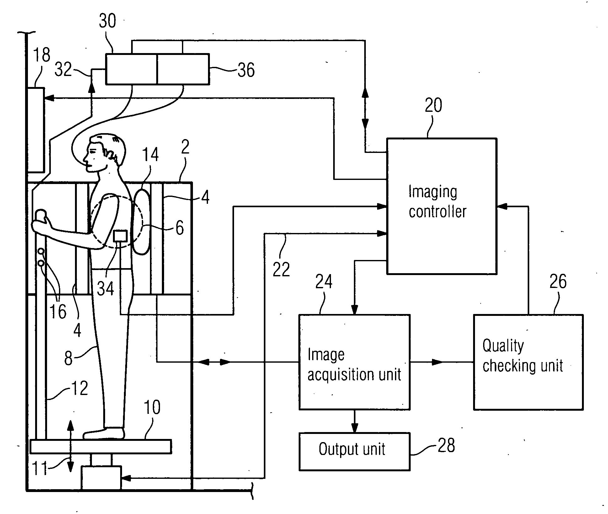
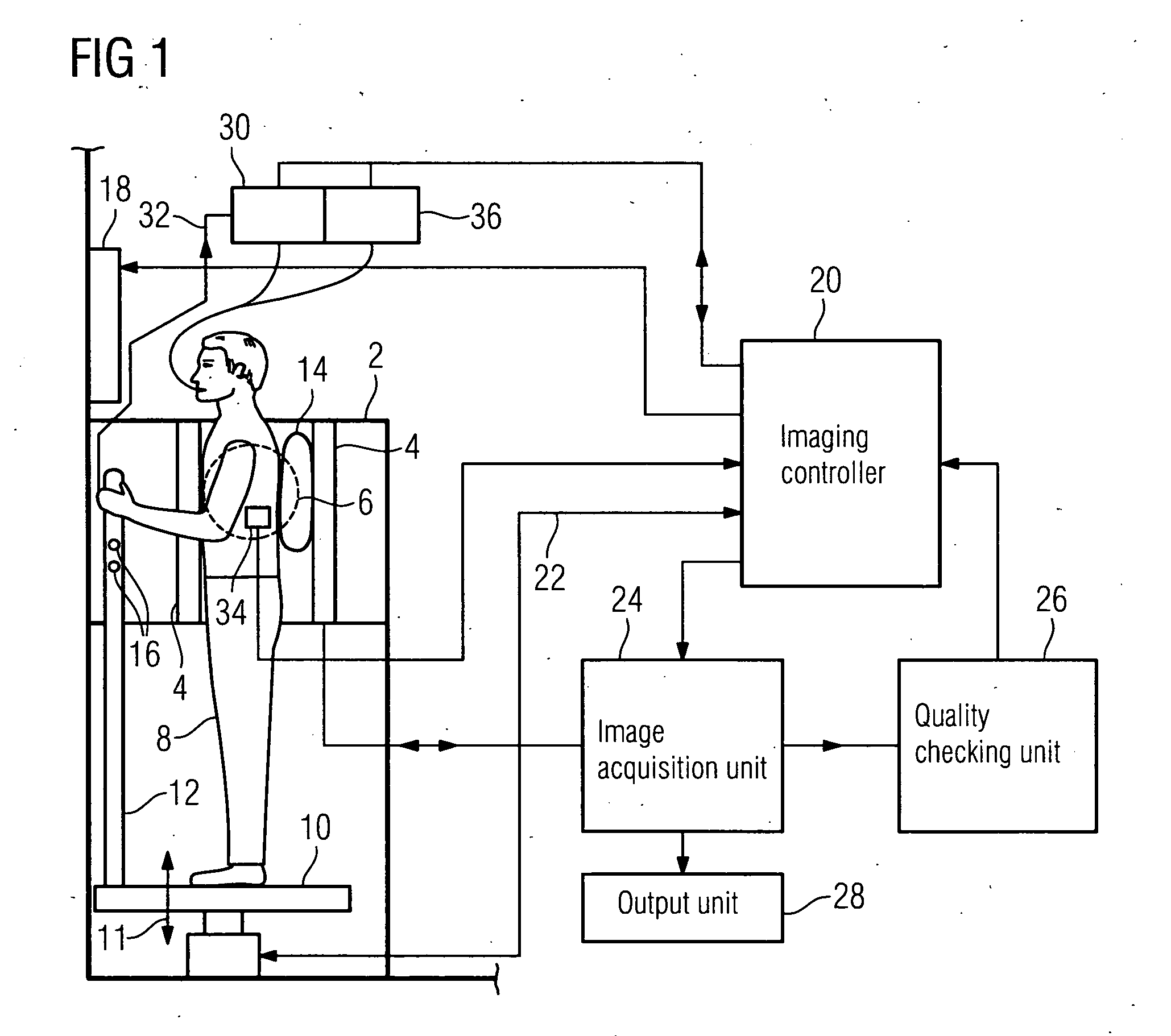
[0038]The upper body MRI scanner shown in FIG. 1 with its main functional units comprises an open magnet arrangement 2 with two oppositely disposed poles 4 between which a homogeneous magnetic field is produced in an imaging volume 6. The magnet poles 4 are interconnected via a magnetic flux return path so as to produce an altogether C-shaped magnet arrangement 2. Possible magnetic field generators include preferably permanent magnet arrangements or even electrically normal-conducting solenoids. The magnetic field strength in the imaging volume is in the order of 0.35 teslas. The magnet arrangement 2 is open to the side so that a patient 8 to be examined can step onto a patient platform 10 supported on the base 6 below the imaging volume 6 in order to assume a standing examination position in the magnet arrangement 2. The patient platform comprises a lifting unit for positioning the patient 8 vertically. The lifting unit is symbolized by the double arrow 11. The upper body MRI scann...


PUM


Abstract

The invention relates to an upper body MRI scanner comprising a magnet arrangement for producing a horizontally oriented homogeneous main magnetic field, said magnet arrangement being designed to examine the upper body of a standing patient. An image acquisition unit is provided for continuously creating upper body images of the standing patient. A quality checking unit is designed for automatically determining a quality metric characterizing the quality of the upper body images. An imaging controller connected to the image acquisition unit and the quality checking unit is provided for activating the image acquisition unit and outputting signals for influencing the position and/or behavior of the patient as a function of the characteristic quality metric. An output unit connected to the image acquisition unit is used for displaying and/or storing at least some of the upper body images.

Description

CROSS REFERENCE TO RELATED APPLICATIONS[0001]This application claims priority of German application No. 10 2007 038 382.9 filed Aug. 14, 2007, which is incorporated by reference herein in its entirety.FIELD OF THE INVENTION[0002]The invention relate to an upper body MRI scanner for examining the upper body of a standing patient. The invention likewise relates to a control method for an upper body MRI scanner of this kind.BACKGROUND OF THE INVENTION[0003]Diagnosing diseases of the cardiopulmonary system is one of the most frequently posed problems in medicine. For example, prior to an operation under anesthetic, investigations must be carried out to determine the patient's cardiopulmonary status. This is done in most cases on the basis of X-ray images. Consequently, the chest X-ray is the most frequent radiological examination of all.[0004]Chest X-ray examination can be used to answer a large number of medical questions with varying degrees of success. This examination is expected to...

Claims


Application Information

Patent Timeline
IPC IPC(8): A61B5/055
CPCA61B5/0555G01R33/281A61B5/0037G01R33/307G01R33/3806G01R33/282A61B5/055A61B5/702A61B5/70
Inventor KUTH, RAINERSCHMIDT, SEBASTIAN
Owner SIEMENS HEALTHCARE GMBH
Why Eureka
Try Eureka