
Biopsy planning system

a biopsy planning and imaging technology, applied in the field of image guided biopsy procedures, can solve the problems that a simple prostate model (e.g., ellipse) with a fixed plan may not be sufficient, and achieve the effects of reducing time during the biopsy procedure, improving workflow, and simple prostate models

Inactive Publication Date: 2009-02-19
EIGEN INC
View PDF54 Cites 26 Cited by
  • Summary
  • Abstract
  • Description
  • Claims
  • Application Information

AI Technical Summary

Benefits of technology

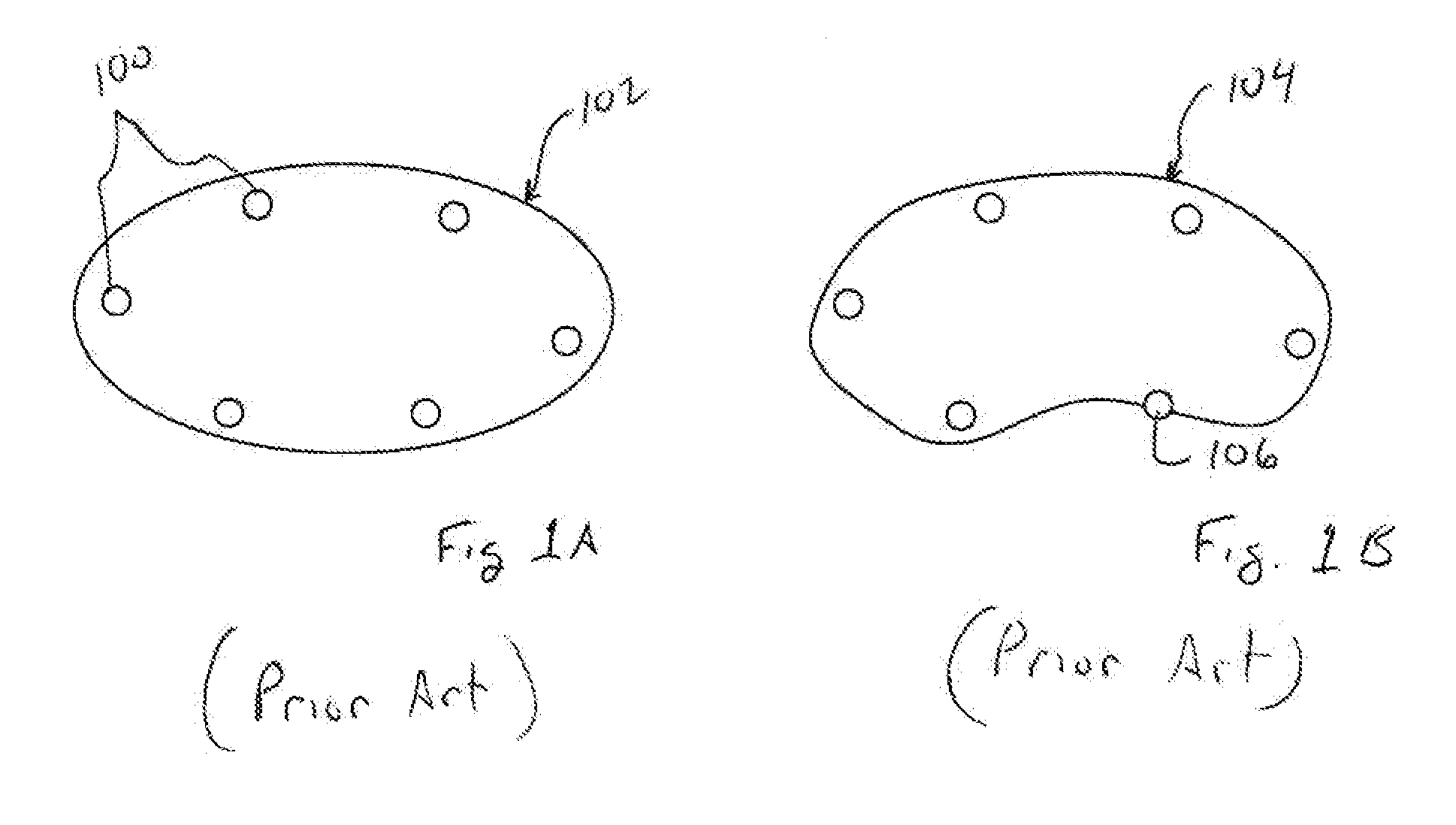
[0009]The utilities allow for automatically loading a standard or a customized biopsy plan onto a medical image. Due to different shapes and sizes of prostates as well as orientation of prostate with respect to the ultrasound probe during image acquisition, a simple prostate model (e.g., ellipse) with a fixed plan may not be sufficient. Accordingly, it has been determined that a deformable shape model with integrated biopsy target locations / sites may be fit to a prostate image. Such a shape model may incorporate standard plans (e.g., sextant, plans, etc.) or customized plans based on, for example, demographic information and / or prostate regions known to be susceptible to cancer (e.g., atlas information and / or previous biopsy information). One main advantage of this utility is full automation (e.g., real time application) of standard or other prepared biopsy plans to a prostate image, which improves workflow and reduces time during the biopsy procedure, while being,accurate. The utility is also flexible to allow the users to also add and refer to the saved customized plans.
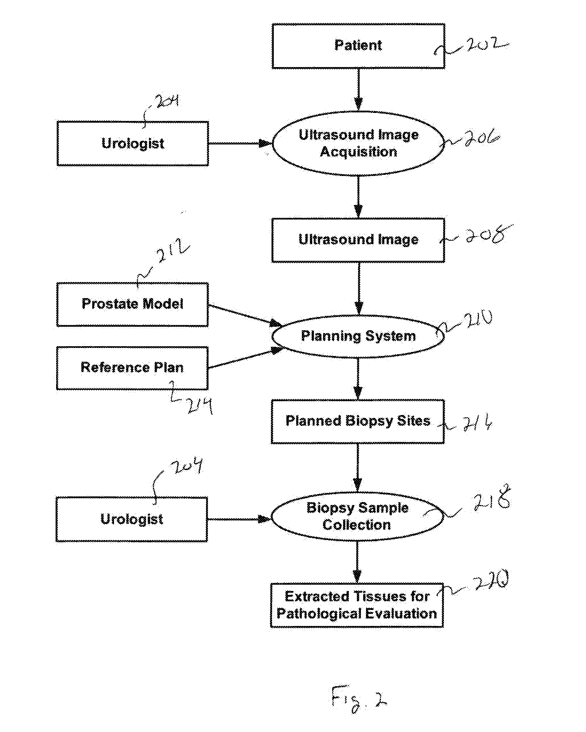
[0010]According to a first aspect, a method for use in applying biopsy target sites to medical prostate images is provided. The method includes obtaining a deformable shape model that is generated from a plurality of prostate images. Use of such a shape model allows for better fitting the model to an acquired prostate image. That is, the shape model, or mean shape of a sample population, allows for better fitting a biopsy plan to different sizes and shapes of prostates as well as orienting the biopsy sites with respect to a currently acquired prostate image. Once the deformable shape model is obtained, a biopsy plan may be identified for use with the shape model. This biopsy plan may then be loaded into the deformable shape model such that the biopsy target locations associated with the plan are registered to locations within the deformable shape model.
[0012]According to another aspect, a method is provided wherein a plurality of biopsy targeting plans are provided to a user during the prostate imaging procedure. The user may then select one or more of the biopsy targeting plans. Accordingly,, biopsy target locations associated with a selected one or more of the biopsy targeting plans may be applied to the image of the prostate. Furthermore, the method includes outputting a processed image of the prostate, with the biopsy target locations illustrated on the processed image. As will be appreciated, this processed image may allow for guidance of a biopsy device to the 1locations of interest (e.g., biopsy target sites).

Problems solved by technology

Due to different shapes and sizes of prostates as well as orientation of prostate with respect to the ultrasound probe during image acquisition, a simple prostate model (e.g., ellipse) with a fixed plan may not be sufficient.

Method used


Image

  • Biopsy planning system
  • Biopsy planning system
  • Biopsy planning system

Examples


Embodiment Construction

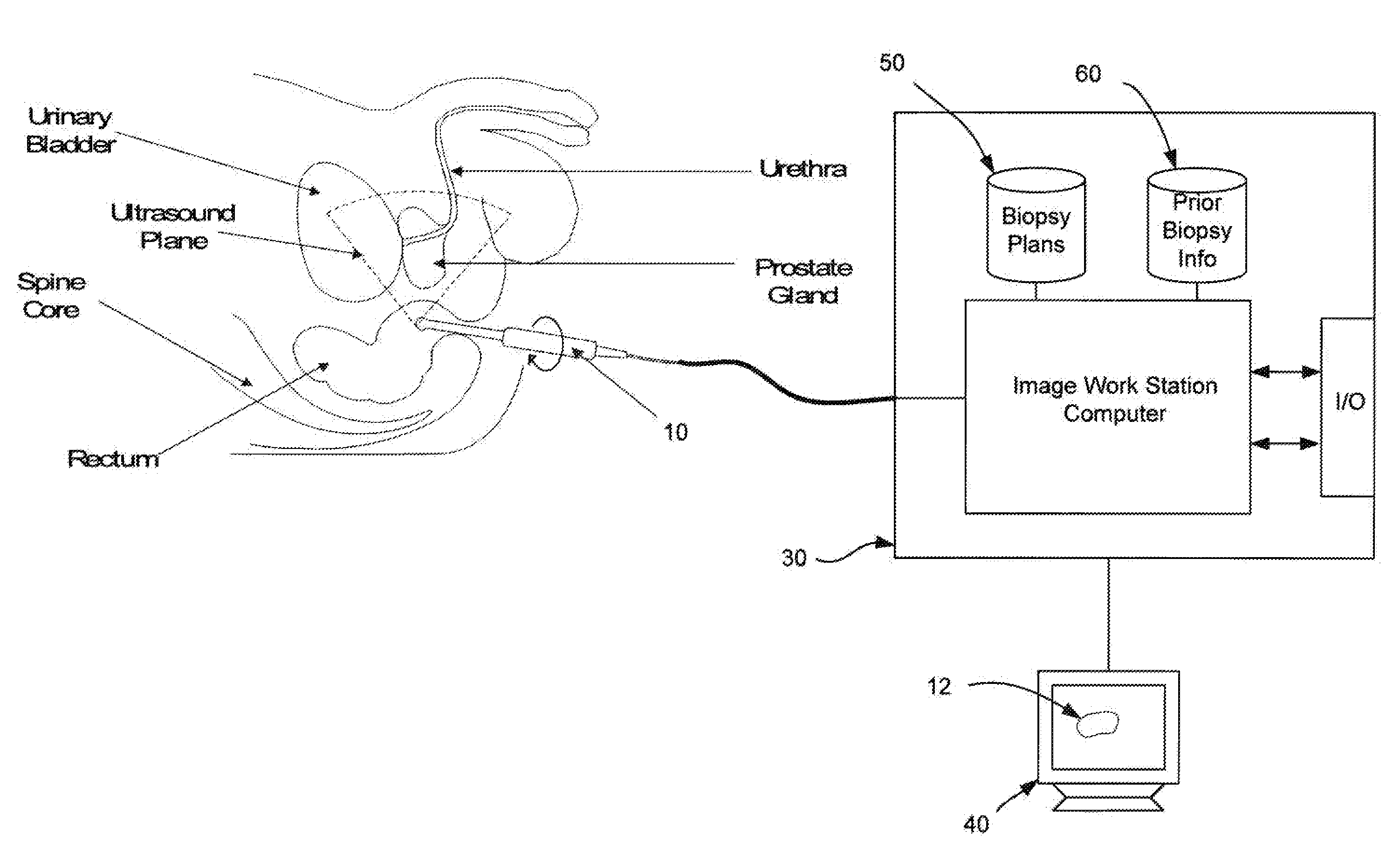
[0029]Reference will now be, made to the accompanying drawings, which assist in illustrating the various pertinent features of the present disclosure. Although the present disclosure is described primarily in conjunction with transrectal ultrasound imaging for prostate imaging it should be expressly understood that aspects of the present invention may be applicable to other,medical imaging applications. In this regard, the following description is presented for purposes of illustration and description.

[0030]Presented herein are systems and processes (utilities) to aid urologists (or other medical personnel) in planning target sites for biopsy. Generally, the utilities use biopsy site model that may be fit (e.g., warped) to an image of a prostate. Such fitting accounts for differently shaped prostates. These, biopsy shape models may incorporate statistical information regarding various zones within a prostate where the cancer resides and / or probability maps of cancer locations obtain...


PUM


Abstract

Guided biopsy is a commonly used method to remove suspicious tissues from an internal organ for pathological tests so that malignancy can be established. Provided herein are systems and methods (i.e., utilities) that allow for automated application of one or more predefined biopsy target plans to an acquired medical image including without limitation, an ultrasound prostate image. Due to different shapes and sizes of prostates as well as orientation of prostate with respect to an ultrasound probe during image, acquisition a simple prostate model (e.g., ellipse) with a fixed plan may not be sufficient. Accordingly, it has been determined that a deformable shape model with integrated biopsy target locations / sites may be fit to a prostate image to provide improved automated biopsy targeting.

Description

FIELD OF INVENTION[0001]The present invention is directed at image guided biopsy procedures. More specifically, the present invention is directed at applying predetermined biopsy locations / plans to medical images to reduce the time required to perform a biopsy procedure and / or improve the accuracy of such a procedure.BACKGROUND OF THE INVENTION[0002]Prostate cancer is the second leading cause of death among males in the USA. However, it, is often curable if detected at an early stage. Accordingly, early detection and treatment is important. In general, a biopsy is recommended when a patient shows high levels of PSA (Prostate Specific Antigen), which is an indicator of prostate cancer or the patient has an abnormal physical exam (e.g., digital rectal exam). Ultrasound has been the main imaging modality for prostate related applications due its inexpensiveness, ease of use, and ability to scan in real-time during biopsy and treatment.[0003]Transrectal Ultrasound (TRUS) Guided Prostate...

Claims


Application Information

Patent Timeline
Patent Type & Authority Applications(United States)
IPC IPC(8): A61B10/02A61B8/13
CPCA61B8/12A61B10/02A61B2019/5295A61B2018/00547A61B2017/00274A61B2090/367
Inventor SURI, JASJIT S.KUMAR, DINESHGUO, YUJUNNARAYANAN, RAMKRISHNAN
Owner EIGEN INC
Learn More