Estimation of Crystal Efficiency With Axially Compressed Sinogram

a sinogram and crystal efficiency technology, applied in tomography, calibration apparatus, instruments, etc., can solve the problems of inaccuracy of crystal efficiency knowledge, poor uniformity of reconstructed image, and higher noise in imag

Active Publication Date: 2009-03-19
SIEMENS MEDICAL SOLUTIONS USA INC
View PDF4 Cites 3 Cited by
  • Summary
  • Abstract
  • Description
  • Claims
  • Application Information

AI Technical Summary

Problems solved by technology

Inaccurate knowledge of crystal efficiency can lead to artifacts, higher noise in the image, and / or poor uniformity in the reconstructed image.
Where, however, there are weaker (i.e. less efficient) detector blocks in a PET system, this approach yields a blurred estimation of crystal efficiency.

Method used


Image

  • Estimation of Crystal Efficiency With Axially Compressed Sinogram
  • Estimation of Crystal Efficiency With Axially Compressed Sinogram
  • Estimation of Crystal Efficiency With Axially Compressed Sinogram

Examples


Embodiment Construction

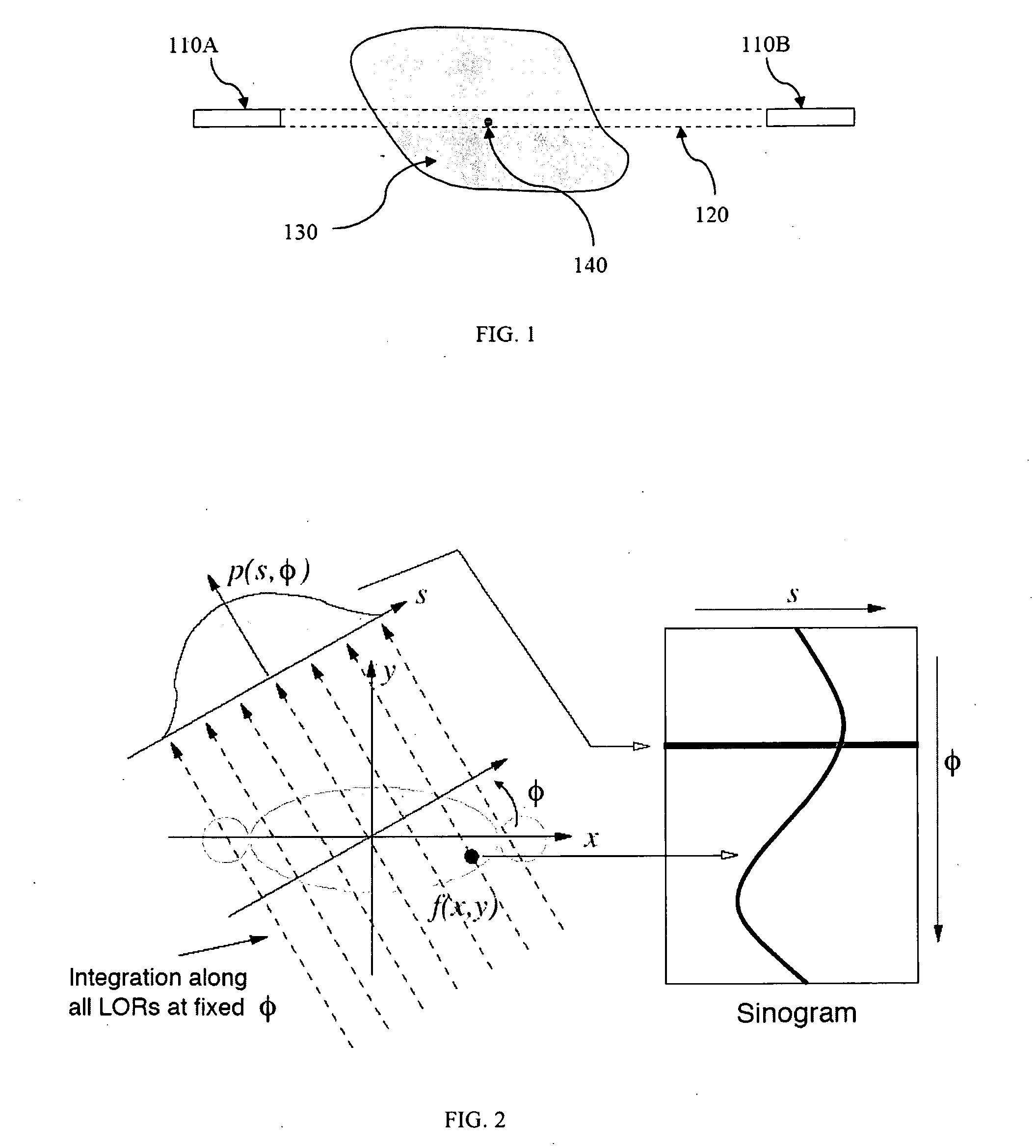
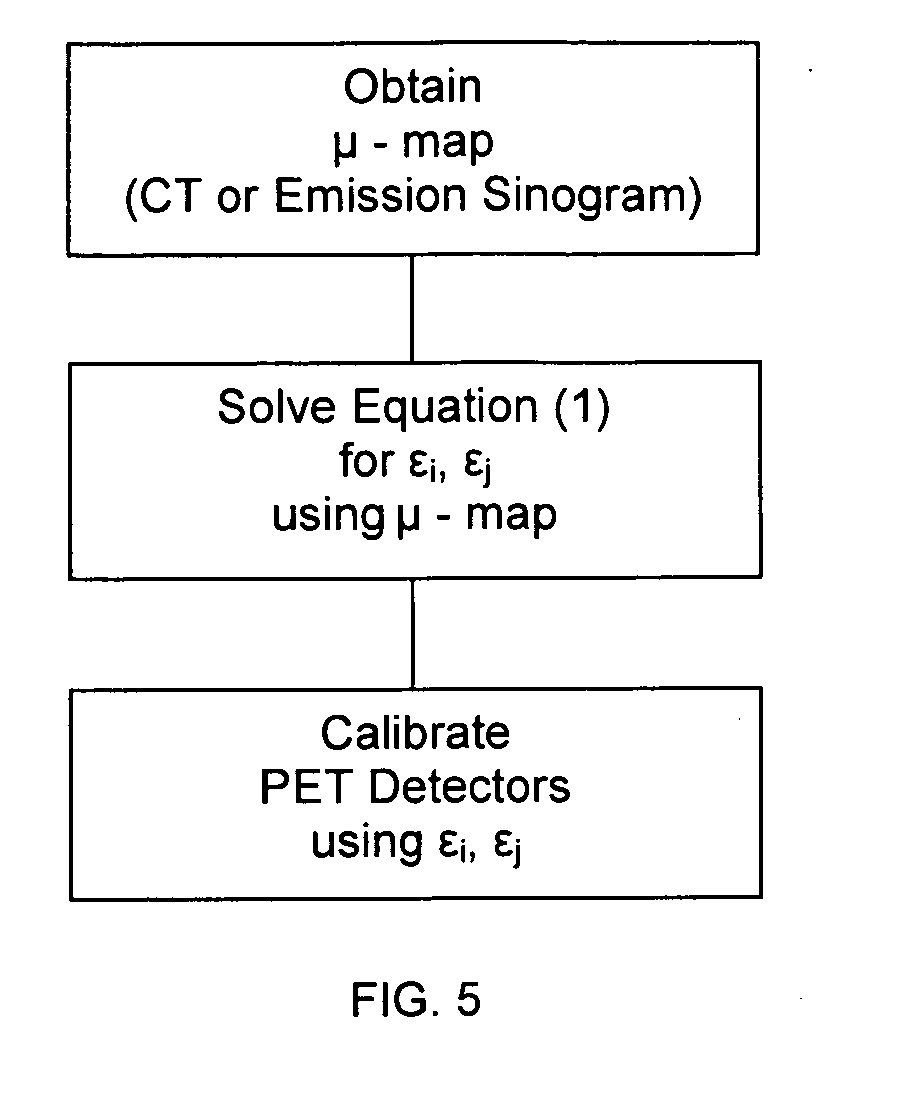
[0022]The present invention enables one to accurately model the contribution to the sinogram bins from each line of response (LOR). To do so, the method utilizes an attenuation correction file generated either from a CT scan or mathematically from an emission sinogram generated using a phantom and iteratively estimates individual crystal efficiency.

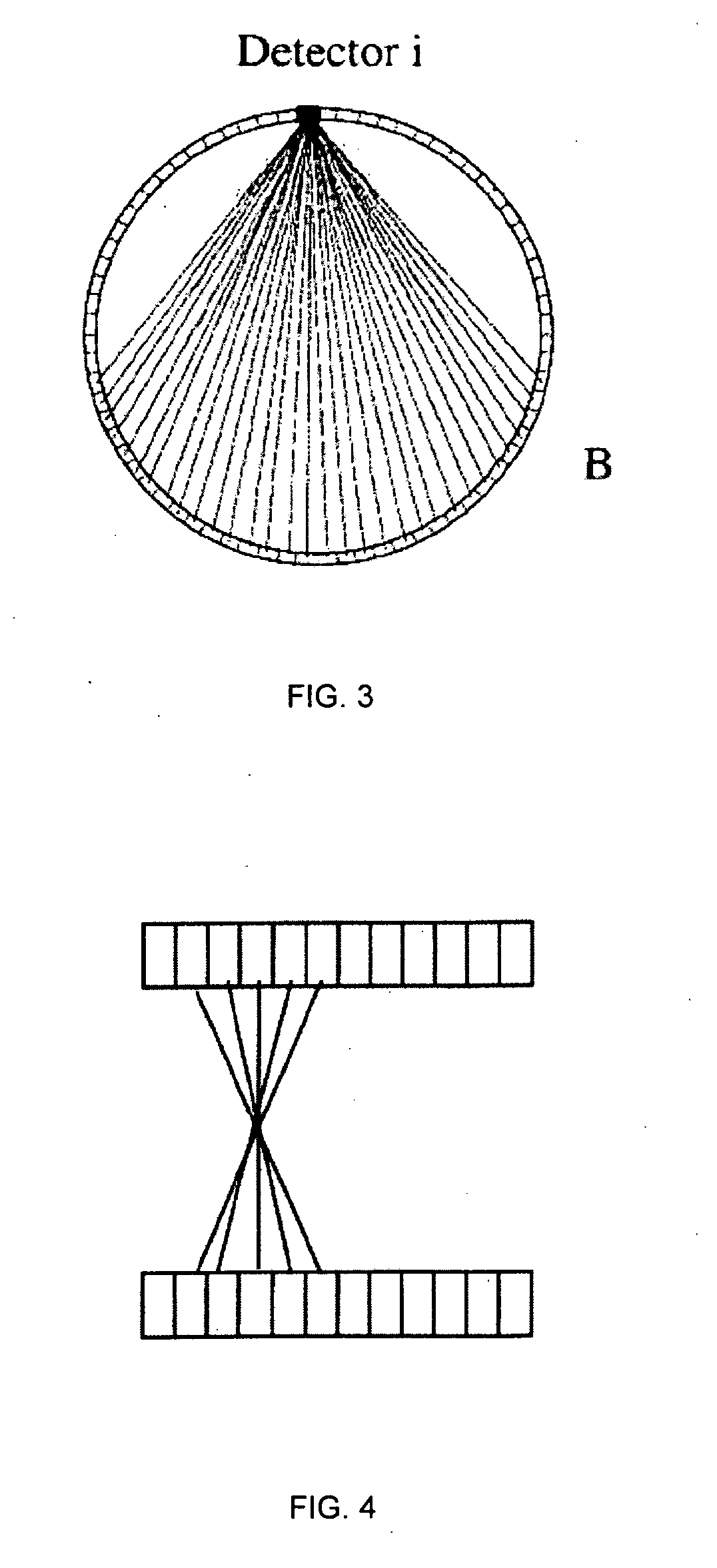
[0023]The fan-sum method is applied on acquired uniform cylinder sinogram to estimate the crystal efficiency of each detector. LORs connecting one crystal i to the opposing group of crystals B are summed together to compute the efficiency of the crystal i, as illustrated in FIG. 3. Thus, in 3D acquisition mode with axial compression, each slice of the sinogram will be the sum of multiple axial lines of response, as illustrated in FIG. 4.

[0024]In the case where all crystal detectors have approximately the same efficiencies, acceptable accuracy can be achieved by ignoring the axial compression and assuming the direct planes are coincidences...


PUM


Abstract

The present invention provides a method for estimating crystal efficiency in a PET detector that takes axial compression into account. It does so via an iterative methodology in which a μ-map is first generated and then is used to obtain a solution for the equationL(ɛi)=∑n∈Nynlog∑i,j∈spangijɛiɛjxij-∑i,j∈spangijɛiɛjxij,wherein gij is a geometric factor for LOR(i,j), εi and εj are the efficiencies for crystal i and crystal j, and xij is the line integral of the source distribution along LOR(i,j). Once efficiencies are determined, they are used to calibrate the PET detector.

Description

CROSS-REFERENCE TO RELATED APPLICATIONS[0001]The present application is based on and claims priority benefit under 35 U.S.C. § 119(e) of provisional application 60 / 973,069 filed Sep. 17, 2007, the contents of which are incorporated by reference.TECHNICAL FIELD[0002]In general, the present invention relates to nuclear medical imaging. More particularly, the invention relates to Positron Emission Tomography (PET) imaging and accurate estimation of scintillation crystal efficiency of PET detector blocks in a PET system.GENERAL BACKGROUND OF THE INVENTION[0003]Nuclear medicine is a unique specialty wherein radiation emission is used to acquire images which show the function and physiology of organs, bones or tissues of the body. The technique of acquiring nuclear medicine images entails first introducing radiopharmaceuticals into the body—either by injection or ingestion. These radiopharmaceuticals are attracted to specific organs, bones, or tissues of interest. Upon arriving at their s...

Claims


Application Information

Patent Timeline
Patent Type & Authority Applications(United States)
IPC IPC(8): G01D18/00
CPCG01T1/2985A61B6/037G01T7/005
Inventor CHEN, MUPANIN, VLADIMIRCASEY, MICHAEL E.
Owner SIEMENS MEDICAL SOLUTIONS USA INC
Why Eureka
Try Eureka