
Method for correcting patient motion when obtaining retina volume using optical coherence tomography

a technology of optical coherence tomography and patient motion, applied in image enhancement, medical/anatomical pattern recognition, instruments, etc., can solve the problem that the method only corrects vertical motion on a b scan, and achieve the effect of eliminating motion artifacts

Active Publication Date: 2009-04-23
OPTOS PLC
View PDF4 Cites 27 Cited by
  • Summary
  • Abstract
  • Description
  • Claims
  • Application Information

AI Technical Summary

Benefits of technology

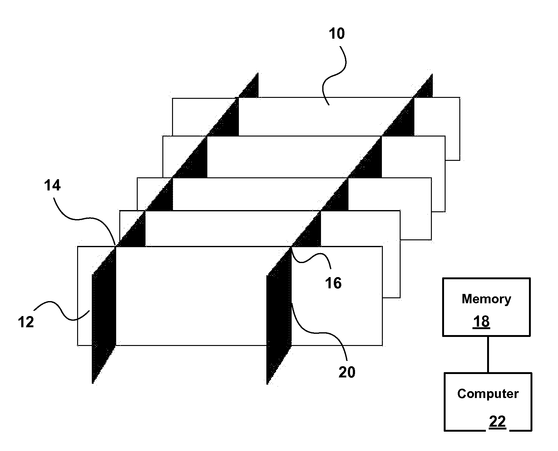
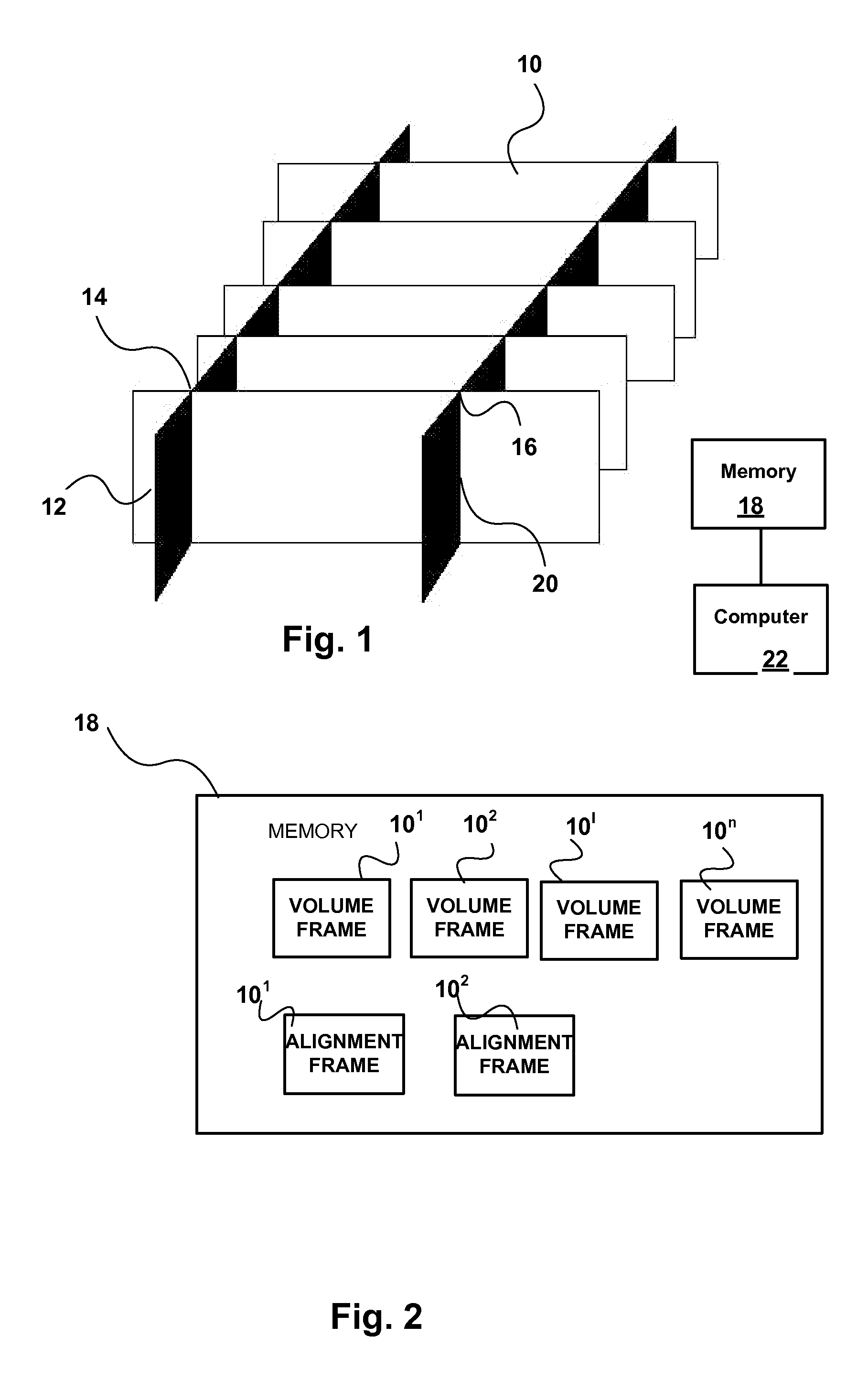
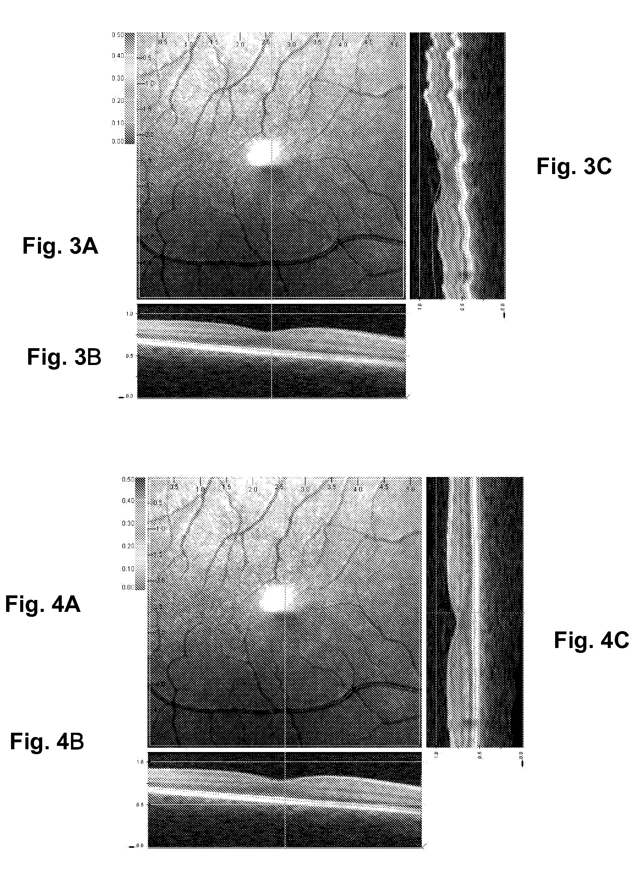
[0007]In accordance with embodiments of the invention, the multiple B-scan images are aligned so that the volume in three dimensions can be reconstructed to look as much like the tissue as possible. The method eliminates motion artifacts caused by patient movement during the capture of OCT volumes. The method will only correct for vertical motion on a B Scan, i.e. the patient moving towards and away from the lens barrel of the OCT scanner.

Problems solved by technology

The method will only correct for vertical motion on a B Scan, i.e. the patient moving towards and away from the lens barrel of the OCT scanner.

Method used


Image

  • Method for correcting patient motion when obtaining retina volume using optical coherence tomography
  • Method for correcting patient motion when obtaining retina volume using optical coherence tomography
  • Method for correcting patient motion when obtaining retina volume using optical coherence tomography

Examples


example

[0026]The following is an algorithm for motion correcting frames assuming that the alignment frames are from ⅓ and ⅔ or the way across each volume frame. The algorithm can be implemented in computer 22.

[0027]The use of 10 column wide sections of image is arbitrary here; any value from 1 to half of the image width could be used:

For each volume frame{ extract 10 columns from the volume frame at ⅓ of it's width smooth the 10 column wide image collapse the 10 column wide image into 1 column extract 10 columns from the ⅓ alignment frame at the relativeposition the above frame is from smooth the 10 column wide image collapse the 10 column wide image into 1 column use statistical methods to determine the best fit for the two images. extract 10 columns from the volume frame at ⅔ of its width smooth the 10 column wide image collapse the 10 column wide image into 1 column extract 10 columns from the ⅔ alignment frame at the relativeposition the above frame is from smooth the 10 column wide im...


PUM


Abstract

A computer-implemented method of correcting for motion of a sample during OCT imaging obtains a series of cross-sectional volume scans through the sample at different positions on a first coordinate axis, obtains at least two cross-sectional alignment scans in planes intersecting said volume scans at an angle, and stores the alignment scans and the volume scans in memory. The alignment scans are matched to the volume scans at lines of intersection thereof to determine the relative displacement of the volume scans to the sample due to sample motion. The relative displacement is used to correct for motion of the sample between successive volume scans.

Description

CROSS REFERENCE TO RELATED APPLICATION[0001]This application claims the benefit under 35 USC 119(e) of prior U.S. provisional application No. 60 / 981,216, the contents of which are herein incorporated by reference.FIELD OF THE INVENTION[0002]This invention relates to the field of optical coherence tomography imaging, and in particular to a method of correcting patient motion while taking scans of the eye.BACKGROUND OF THE INVENTION[0003]Optical coherence tomography (OCT) is an imaging technique that makes possible the three dimensional imaging of transparent or semi-transparent objects, such body tissues, specifically eye. OCT is described in detail in U.S. Pat. No. 6,769,769, the contents of which are herein incorporated by reference.[0004]Using Optical Coherence Tomography (OCT) it is possible to rapidly obtain a line of image information extending in the z direction, where the z axis extends in the depth direction. Such a scan is known as an A-scan. In time domain OCT, this can be...

Claims


Application Information

Patent Timeline
Patent Type & Authority Applications(United States)
IPC IPC(8): A61B3/14G06K9/00
CPCA61B3/102A61B3/1225G06K9/6203G06T2207/30041G06T7/0024G06T2207/10101G06K2209/05G06T7/30G06V10/7515G06V2201/03
Inventor MCLEAN, DUNCANPEDRO, JUSTINHAYWARD, CLIVE
Owner OPTOS PLC
Learn More