Interactive Medical Imaging Processing and User Interface System

a medical imaging and user interface technology, applied in the field of interactive medical imaging processing and user interface system, can solve the problems of time-consuming, labor-intensive and burdensome, and substantial work burden involving difficult ventricular angiogram processing

Inactive Publication Date: 2009-04-23
SIEMENS MEDICAL SOLUTIONS USA INC
View PDF25 Cites 15 Cited by
  • Summary
  • Abstract
  • Description
  • Claims
  • Application Information

AI Technical Summary

Problems solved by technology

This known process is time consuming, labor intensive and burdensome and average ED and ES selection time typically comprises more than 1 minute and involves 40 to 50 user selection commands for one patient imaging study.
Many (e.g., hundreds) of left ventricular analyses may need to be performed daily in a catheterization department representing a substantial work burden involving difficult ventricular angiogram processing.

Method used


Image

  • Interactive Medical Imaging Processing and User Interface System
  • Interactive Medical Imaging Processing and User Interface System
  • Interactive Medical Imaging Processing and User Interface System

Examples


Embodiment Construction

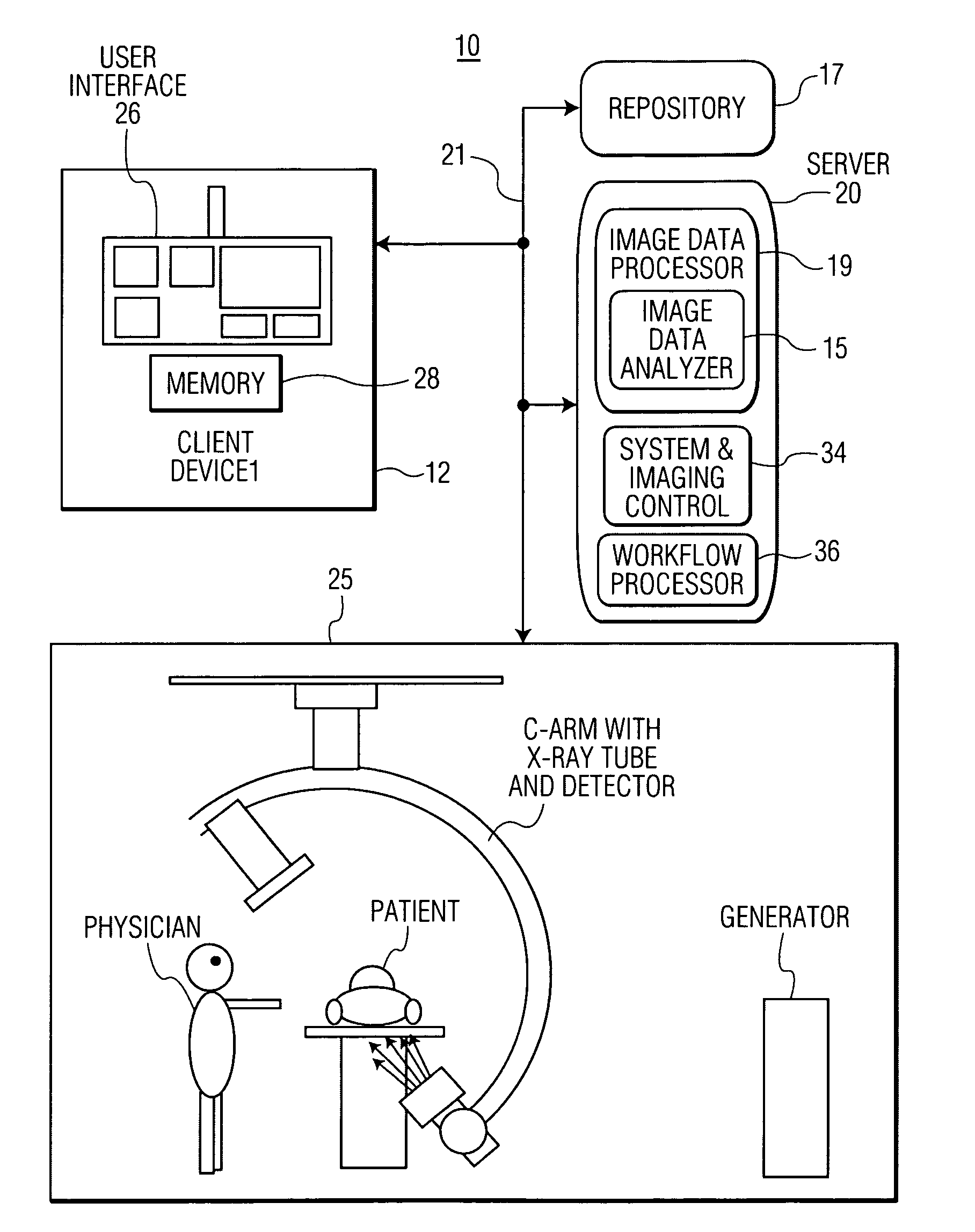
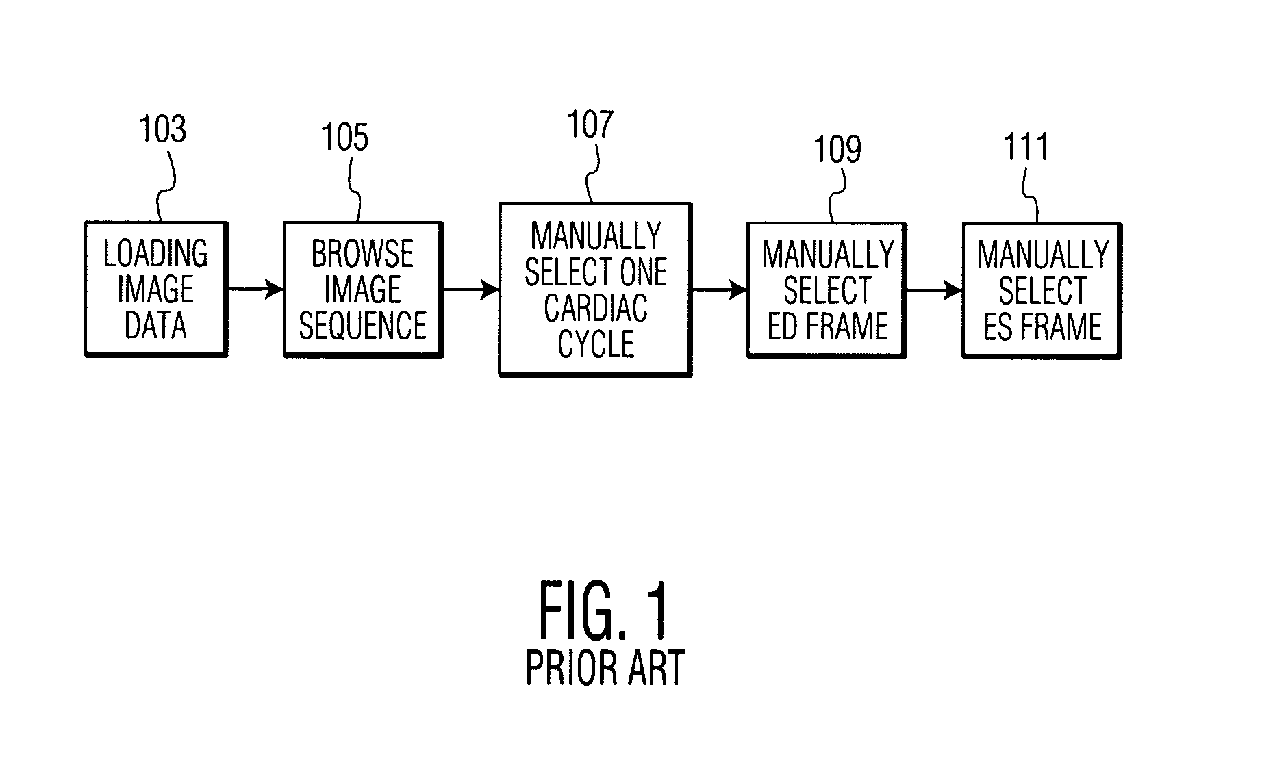
[0011]An interactive medical image processing and user interface system presents a user interactive image window including a distribution curve of an organ section area over a heart beat cycle time and an image of a patient organ and supports a desired clinical workflow. FIG. 3 illustrates a clinical workflow employed by the interactive medical image processing and user interface system in left ventricular analysis, for example. In the workflow process the system acquires and loads data representing multiple images of an organ of a patient in step 303 and in steps 305 and 307 automatically detects an end-diastolic image frame (ED) and end-systolic image frame (ES) using one of a variety of different known processes and provide frame numbers indicating the identified images. The ED and ES images are thereby subsequently accessible for display on a workstation. In step 309, the system also estimates a distribution of patient left ventricle area change over multiple heart cycles for di...


PUM


Abstract

An interactive medical user interface system presents a user interactive image window, including a distribution curve of an organ section area over a heart beat cycle time and supports a desired clinical workflow. An interactive medical image processing and user interface system for use in patient organ imaging includes an image data processor. The image data processor processes data representing multiple images of an organ of a patient over the heart beat cycle of the patient to derive data representing a distribution curve of an organ section area over a heart beat cycle time. A user interface generates data representing a composite user interface display image including, a first user interactive image window presenting the distribution curve and a second image window presenting an image of the organ corresponding to a location on the distribution curve interactively selected by a user via the first user interactive image window.

Description

[0001]This is a non-provisional application of provisional application Ser. No. 60 / 981,222 filed Oct. 19, 2007, by W. Qu.FIELD OF THE INVENTION [0002]This invention concerns an interactive medical image processing and user interface system for use in patient anatomical organ imaging involving presenting a user interactive image window including a distribution curve of an organ section area over a heart beat cycle time and an image of a patient organ.BACKGROUND OF THE INVENTION [0003]Ventricular angiography is commonly employed in examining cardiac functions and determining heart parameters representing stroke volume, ejection fraction, and heart wall motions. Known systems involve determining an end-diastolic image frame (ED) and an end-systolic image frame (ES) from an angiographic image sequence for use in quantizing heart parameters. Known systems typically employ a workflow for ED and ES selection in left ventricular analysis as illustrated in FIG. 1. In the known clinical workf...

Claims


Application Information

Patent Timeline
Patent Type & Authority Applications(United States)
IPC IPC(8): A61B5/05G16H30/40G16H40/63
CPCA61B6/4441G06F19/3406G06F19/321A61B6/463G16H40/63G16H30/40
Inventor QU, WEI
Owner SIEMENS MEDICAL SOLUTIONS USA INC
Why Eureka
Try Eureka