Nuclear medicine diagnosis device, form tomography diagnosis device, nuclear medicine data arithmetic processing method, and form tomogram arithmetic processing method

a nuclear medicine and diagnostic device technology, applied in tomography, x/gamma/cosmic radiation measurement, instruments, etc., can solve the problems of deteriorating profile extraction accuracy, inability to secure profiles, and inability to extract profiles, so as to improve the accuracy of profile extraction, improve the accuracy of absorption coefficient maps, and improve the accuracy of absorption correction

Inactive Publication Date: 2009-07-02
SHIMADZU CORP
View PDF1 Cites 32 Cited by
  • Summary
  • Abstract
  • Description
  • Claims
  • Application Information

AI Technical Summary

Benefits of technology

[0049]As can be seen, in case of the latter map, a more accurate absorption coefficient map can be created according to an actual subject and more accurate absorption correction can be thereby conducted.
[0050]Moreover, the absorption coefficient map is not necessarily used...

Problems solved by technology

Nevertheless, if the detectors are located to be proximate to the subject with views of improving sensitivity and spatial resolution, a space for mounting a collimated external radiant source, a mechanism that rotates the radiant source (radiant source rotation mechanism) and the like is not often secured.
However, if radioactive accumulation is quite small on edges of the subject, the...

Method used


Image

  • Nuclear medicine diagnosis device, form tomography diagnosis device, nuclear medicine data arithmetic processing method, and form tomogram arithmetic processing method
  • Nuclear medicine diagnosis device, form tomography diagnosis device, nuclear medicine data arithmetic processing method, and form tomogram arithmetic processing method
  • Nuclear medicine diagnosis device, form tomography diagnosis device, nuclear medicine data arithmetic processing method, and form tomogram arithmetic processing method

Examples


first embodiment

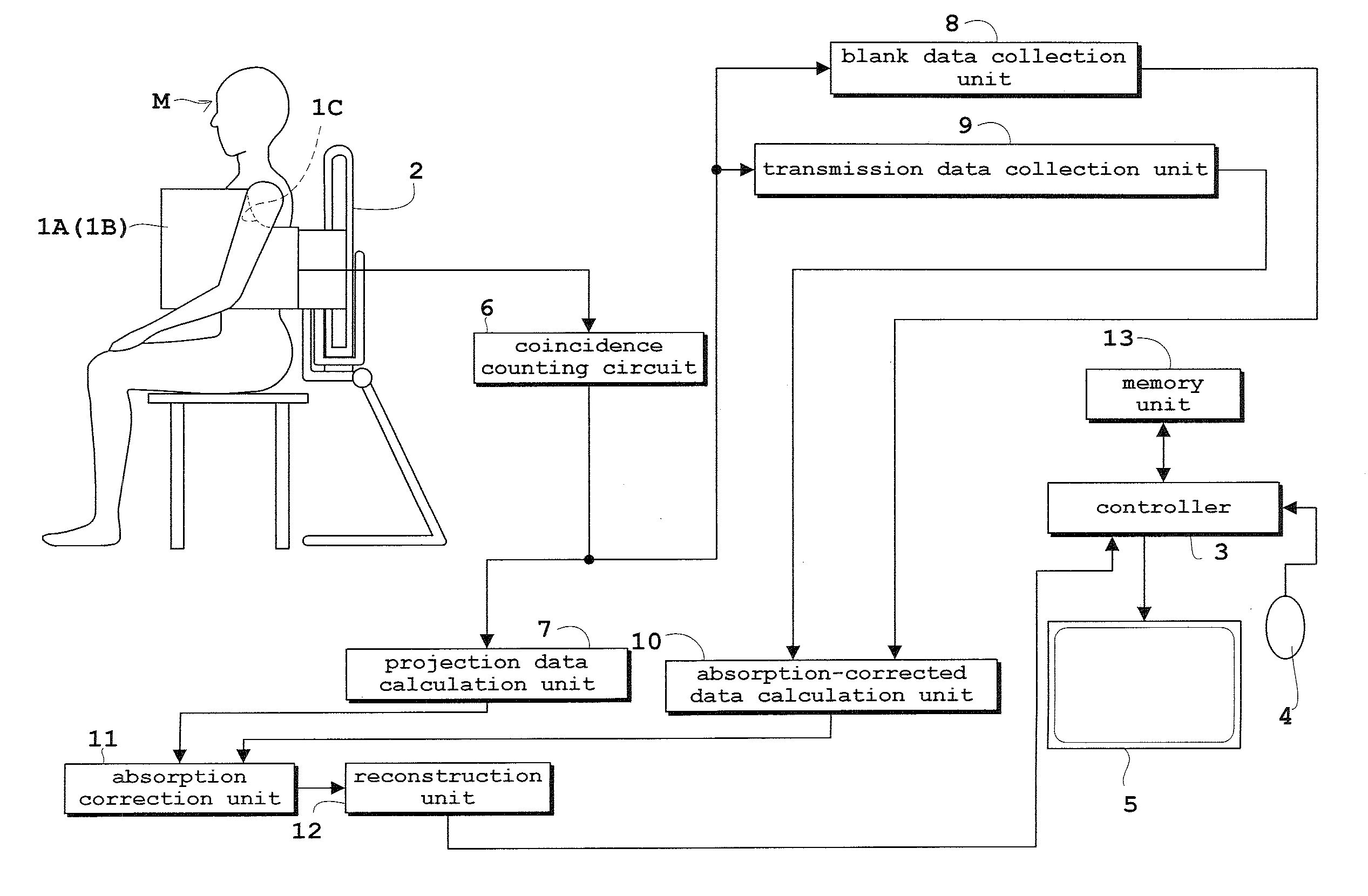
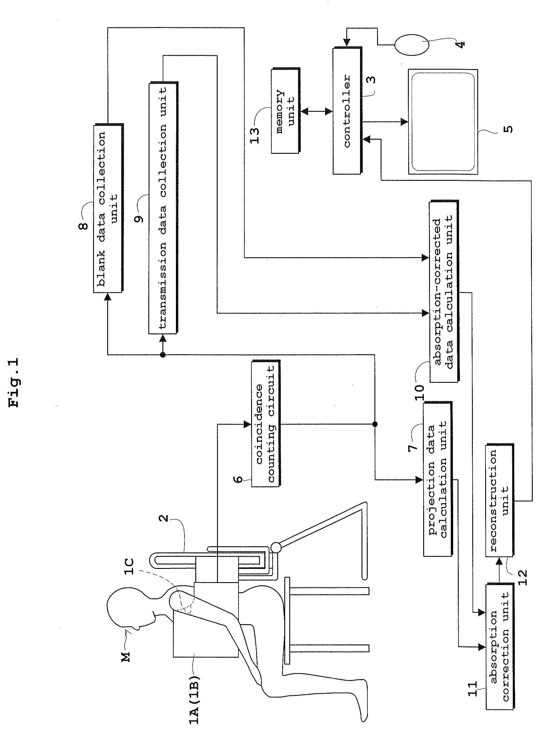
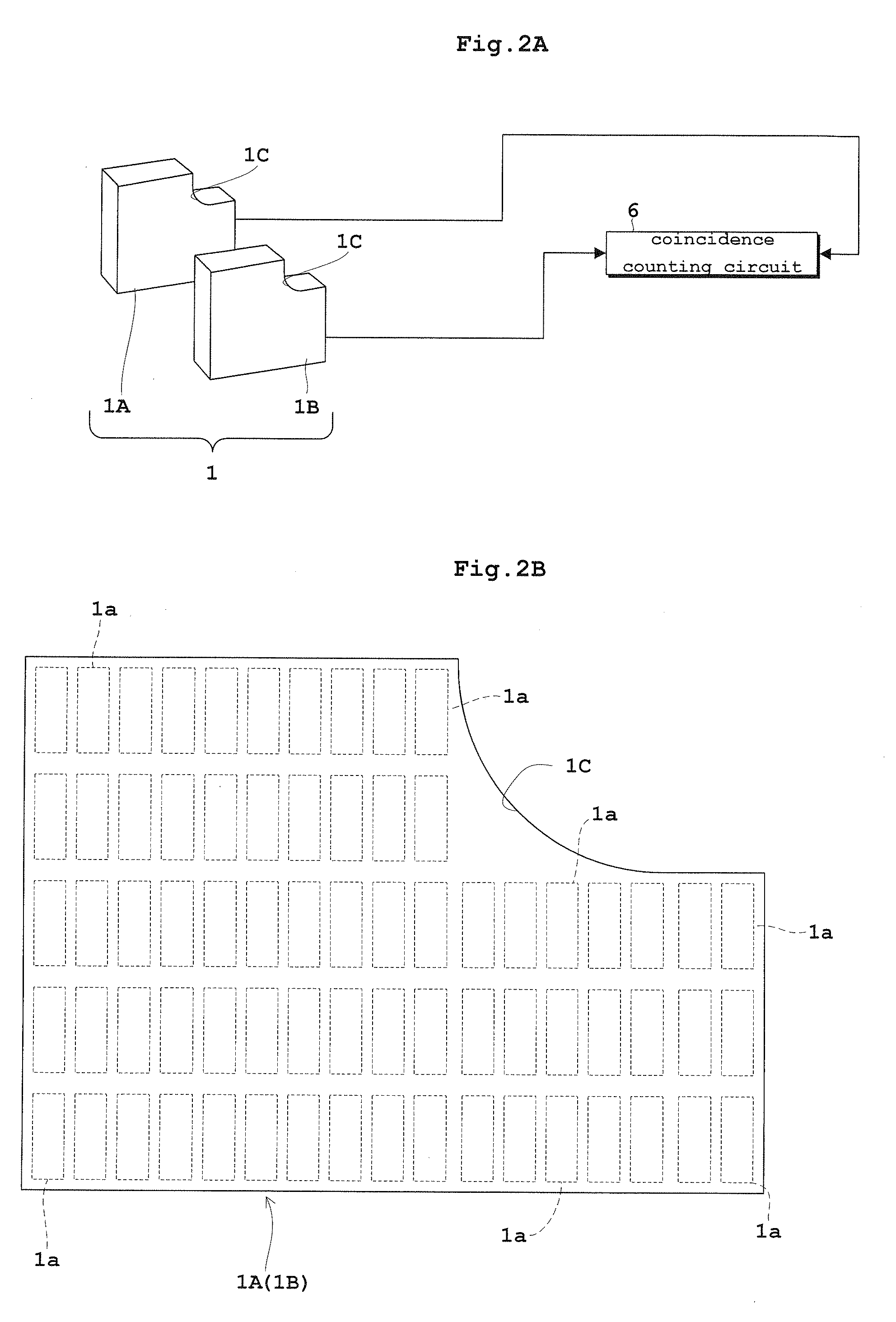
[0073]FIG. 1 is a side view and a block diagram of a positron emission tomography (PET) mammography device according to a first embodiment. FIGS. 2A and 2B are a block diagram showing surroundings of detector plates used in the PET mammography device according to the first embodiment and a schematic diagram of each of the detector plates, respectively. FIG. 3 is schematic side view showing a specific configuration of a radiation detector in each detector plate. FIGS. 4A and 4B are schematic diagrams showing modes of scintillators constituting each radiation detector, respectively. In the first embodiment as well as second and third embodiments to be described later, a PET device will be described by way of example as a nuclear medicine diagnosis device. In the first embodiment, the PET mammography device applied to mammograms for breast cancer detection will be described as an example of the PET device.

[0074]As shown in the block diagrams of FIGS. 1 and 2A, the PET mammography devic...

second embodiment

[0117]A second embodiment of the present invention will be described below with reference to the drawings.

[0118]FIG. 7 is a side view and a block diagram of a PET device according to the second embodiment. FIG. 8 is a schematic diagram of a ring radiation detection mechanism employed in the PET device according to the second embodiment. In the second embodiment, similarly to the first embodiment, the PET device will be described as a nuclear medicine diagnosis device by way of example. In the second embodiment, the PET device including ring radiation detection mechanisms 1D downsizing of which is realized by making the mechanisms 1D as proximate to the subject M as possible except for the external radiant source will be described by way of example.

[0119]As shown in FIG. 7, the PET device according to the second embodiment includes a controller 3, an input unit 4, an output unit 5, a coincidence counting circuit 6, a projection data calculation unit 7, a blank data collection unit 8,...

third embodiment

[0150]A third embodiment according to the present invention will be described below with reference to the drawings.

[0151]FIG. 13 is a side view and a block diagram of a PET mammography device according to the third embodiment. FIG. 14 is a side view and a block diagram of a PET device according to the third embodiment. In the third embodiment, similarly to the preceding first and second embodiments, a form tomography diagnosis device will be described while taking the PET mammography device and the PET device as examples. In the third embodiment, an instance in which the present invention is applied to a PET mammography device similar to that according to the first embodiment and an instance in which the present invention is applied to a PET device including ring radiation detectors 1D similar to that according to the second embodiment will be described with reference to FIG. 13 showing the side view and the block diagram of the PET mammography device and to FIG. 14 showing the side...


PUM


Abstract

In a state in which a subject is absent, blank data is collected by a self-radioactivity element typified by Lu-176 (S1). In a state in which the subject is present, transmission data is collected by the self-radioactivity element (S2). Emission data is collected by γ rays emitted from the subject injected with a radiopharmaceutical (S3). Absorption-corrected data is calculated based on the blank data and the transmission data (S4 to S7), and the emission data is absorption-corrected using the absorption-corrected data (S8). Although such background data obtained by the self-radioactivity is originally abandoned, the background data is rather used for the absorption-corrected data. Stable absorption correction can be thereby conducted.

Description

BACKGROUND OF THE INVENTION[0001](1) Field of the Invention[0002]The present invention relates to a nuclear medicine diagnosis device, a form tomography diagnosis device, a nuclear medicine data arithmetic processing method, and a form tomogram arithmetic processing method for obtaining nuclear medicine data or form tomograms of a subject injected with radiopharmaceutical based on radiant rays generated from the subject.[0003](2) Description of the Related Art[0004]The nuclear medicine diagnosis device, that is, an ECT (Emission Computed Tomography) device will be described while taking a PET (Positron Emission Tomography) device as an example. The PET device is configured to detect a plurality of gamma (γ) rays generated as a result of annihilation of positrons and to reconstruct tomograms of a subject only when a plurality of detectors detects the γ rays simultaneously.[0005]Specifically, a radiopharmaceutical containing a positron-emitting radionuclide is administered into the su...

Claims


Application Information

Patent Timeline
IPC IPC(8): G06K9/00
CPCG01T1/1612A61B6/037G01T1/1617A61B6/00
Inventor MIZUTA, TETSUROKITAMURA, KEISHI
Owner SHIMADZU CORP
Why Eureka
Try Eureka