Method and Devices for Minimally Invasive Arthroscopic Procedures

a minimally invasive and arthroscopic technology, applied in the field of arthroscopic procedures, can solve the problems of inability to access the posterior side of the joint, the damage to the structure is permanent and irreparable, and the risk of the procedure is significantly increased

Active Publication Date: 2009-07-02
ORTHODYNAMIX
View PDF52 Cites 67 Cited by
  • Summary
  • Abstract
  • Description
  • Claims
  • Application Information

AI Technical Summary

Benefits of technology

[0021]In another aspect, the invention generally relates to a method of performing minimally invasive diagnostic and surgical procedures on the hip comprising (a) providing a visualization and / or an operable device(s) comprising a handle at a proximal end; an operable end at a distal end; a rigid or semi rigid elongate body member extending between the handle and the operable end; a distal flexible end segment that rotatably connects the operable end to the elongate body member; the handle comprising control means to precisely maneuver the operable end by iteratively adjusting each of the following degrees of freedom: linear translation of the operable end into the hip joint capsule; rotation about the linear axis of the elongated body member; curvilinear bending of the distal flexible end segment; and rotation about an axis of a bend in the distal end segment; (b) disposing the flexible end segment into a straight configuration; (c) inserting the distal end of the device into the body and into the hip capsule; (d) linearly translating the operable end into the capsule; (e) iteratively adjusting the curvilinear bend radius of the distal flexible end segment while translating the operable end toward the operative target; (f) performing the procedure; (g) disposing the end segment into a straight configuration; and (h) removing the device from the capsule.

Problems solved by technology

Damage to these structures is permanent and irreparable
Such an added portal considerably increases the risk of the procedure because the proximity of the lateral femoral cutaneous nerve, the lateral femoral circumflex artery, and the femoral neurovascular bundle.
Access via the opposite, posterior side of the joint, i.e. the gluteal region, is not a viable option nor is the medial approach from the groin.
While the situation can be relieved somewhat through the use of 70 degree scopes and physically prying the cannulas into a contrived position, the access problem remains a significant hurdle to the performance of arthroscopic procedures on the hip.

Method used


Image

  • Method and Devices for Minimally Invasive Arthroscopic Procedures
  • Method and Devices for Minimally Invasive Arthroscopic Procedures
  • Method and Devices for Minimally Invasive Arthroscopic Procedures

Examples


Embodiment Construction

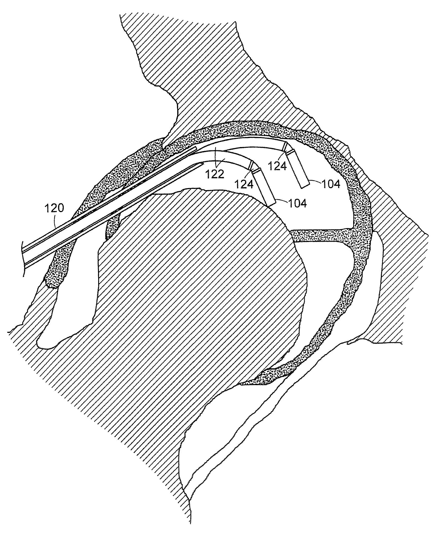
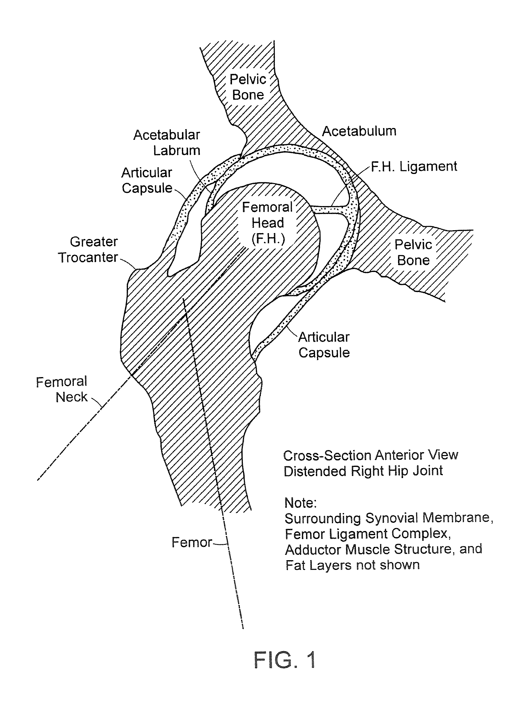
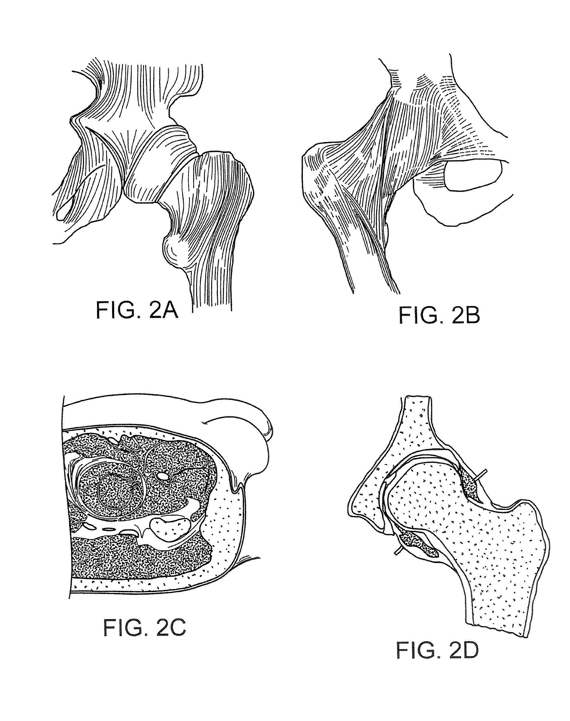
[0072]The devices and methods of the invention are primarily illustrated and described herein by means of devices which have been adapted for use in performing arthroscopic procedures on the hip. The devices and methods provide access to the internal portions of the distended hip capsule during arthroscopic procedures that are presently not accessible using currently available arthroscopic instruments. The devices and methods can suitably be used to perform arthroscopic procedures not only on the hip, but also on other parts of the body, such as the knee and shoulder. The devices are particularly suitable for performing procedures on parts of the body that require flexible access. The devices and methods are not limited to arthroscopy, and can further be used in endoscopic and laparoscopic procedures as well as open surgeries. The devices can be in the general form of any conventional diagnostic or operative instrument including, but not limited to, gaspers, scissors, forceps, scalp...


PUM


Abstract

A surgical device is provided, the device having a tubular outer body member with a handle at its proximal end and a flexible distal end segment extending from its distal end. The surgical device also has an operable end extending from the distal end of the flexible distal end segment. The device is configured for selectively causing the flexible end segment to bend to adopt a desired curvature and for selectively rotating the operable end relative to the flexible distal end segment about an axis of the flexible distal end segment.

Description

CROSS-REFERENCE TO RELATED APPLICATIONS[0001]The present application is a continuation-in-part of U.S. patent application Ser. No. 12 / 119,799, filed on May 13, 2008, which is a continuation of U.S. patent application Ser. No. 11 / 643,740, filed Dec. 20, 2006, which claims the benefit of U.S. provisional application 60 / 752,284, filed Dec. 20, 2005, each of which is incorporated herein by reference in its entirety.FIELD OF THE INVENTION[0002]The present invention relates generally to devices and methods for performing arthroscopic procedures, particularly arthroscopic procedures on the hip, including arthroscopic diagnostic and surgical procedures.BACKGROUND OF THE INVENTION[0003]Access to the knee and shoulder capsules during arthroscopic surgery is typically made through opposing portals often called the operative portal and the visualization portal. The arthroscope is typically inserted through the visualization portal, while the medical device is inserted through the operative port...

Claims


Application Information

Patent Timeline
Patent Type & Authority Applications(United States)
IPC IPC(8): A61B1/00
CPCA61B1/00165A61B2017/2906A61B1/05A61B1/317A61B17/29A61B18/1482A61B2017/003A61B2017/00867A61B2017/2904A61B2017/2905A61B2017/2931A61B2017/2948A61B1/00147A61B17/320016A61B18/1492A61B19/22A61B2017/2903A61B2019/2242A61B17/295A61B2017/00331A61B1/0055A61B34/70A61B34/71A61B2017/291
Inventor JORGENSEN, GLENDENNIS, WILLIAM G.
Owner ORTHODYNAMIX
Why Eureka
Try Eureka