Method and apparatus for coordinating contrast agent injection and image acquisition in c-arm computed tomography

Inactive Publication Date: 2009-08-06
SIEMENS AG
View PDF7 Cites 115 Cited by
  • Summary
  • Abstract
  • Description
  • Claims
  • Application Information

AI Technical Summary

Benefits of technology

[0012]An object of the present invention is to improve the manner by which the aforementioned timing between contrast agent injection and image acquisition is determined or controlled, in the context of computed tomography. The above object is achieved in accordance with the present invention by a method and an apparatus wherein the injection of the contrast agent is monitored by operating the computed tomography apparatus in a radiographic (fluoroscopic) mode, followed by image acquisition with the same CT apparatus being operated in the

Problems solved by technology

Since many factors can enter into the progress of the injected contrast agent through the vascular system of the examination subject, it is difficult to accurately predict the point in time at which the contrast agent bolus will arrive at, and optimally fill, the vessels in the region of interest, for which the three-dimensional image is to be reconstructed.
In practice, however, the predetermined timing that is embod

Method used


Image

  • Method and apparatus for coordinating contrast agent injection and image acquisition in c-arm computed tomography
  • Method and apparatus for coordinating contrast agent injection and image acquisition in c-arm computed tomography

Examples


Example

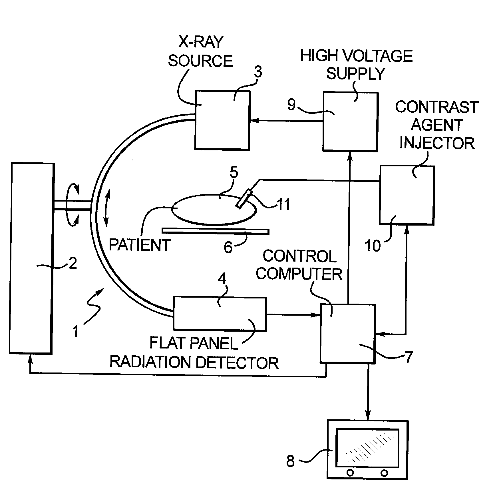
[0019]The invention is explained in the context of the embodiment shown in the figure, on the basis of a C-arm apparatus 1. The employment of the invention, however, is not limited to a C-arm apparatus. The principles of the invention can also be embodied in a “classic” CT apparatus of the type described in U.S. Pat. No. 6,487,267, having a rotating gantry mounted in a stationary frame.

[0020]In the embodiment shown in the figure, the C-arm apparatus 1 has a C-arm stand 2 that supports an x-ray source 3 and a flat panel radiation detector 4 so as to be rotatable around a patient 5 on a patient bed 6. Such rotational capability is indicated by the circular double arrow. The C-arm apparatus 1 is also capable of orbital movement, as indicated by the curved double arrow, as well as conventional vertical movement (not separately indicated).

[0021]The C-arm apparatus 1 is operated by a control computer 7, which supplies movement signals to the C-arm stand 2 to operate motors or other moveme...


PUM


Abstract

In a medical imaging system and method, an image data acquisition system is operable in a fluoroscopy mode to obtain a fluoroscopic image of a subject and is operable in a CT mode to obtain projection data sets of the subject that are used to reconstruct a three-dimensional image of a region of interest of the subject. For imaging procedures involving the administration of contrast agent to the subject, the image data acquisition apparatus is operated in the fluoroscopy mode at the time the administration of contrast agent is begun, and the filling of vessels with the contrast agent is automatically monitored by fluoroscopy. When the automatic monitoring indicates that an optimal degree of filling of the vessels with the contrast agent has occurred, the image data acquisition apparatus is automatically switched to operate in the CT mode to acquire projection data sets of the region of interest containing the vessels filled with contrast agent.

Description

BACKGROUND OF THE INVENTION[0001]1. Field of the Invention[0002]The present invention relates to C-arm computed tomography, and in particular to C-arm computed tomography for the purpose of generating angiographic images of an examination subject, with contrast agent being administered to the subject.[0003]2. Description of the Prior Art[0004]C-arm computed tomography is being increasingly used for the purpose of acquiring and depicting three-dimensional structures in a patient immediately before or during a minimally invasive medical interventional procedure.[0005]The anatomical structures of interest are often vessels or other body lumens, the depiction of which in the reconstructed image requires the administration (injection) of a contrast agent to the examination subject. For this purpose, it must be ensured that, during the image acquisition, the contrast agent is present with a sufficiently high concentration in the region of the volume of interest for which three-dimensional...

Claims


Application Information

Patent Timeline
IPC IPC(8): A61B5/05
CPCA61B6/03A61B6/504A61B6/481A61B6/4441
Inventor HOHEISEL, MARTIN
Owner SIEMENS AG
Why Eureka
Try Eureka