Tagged Ligands For Enrichment of Rare Analytes From A Mixed Sample

a technology of rare analytes and tagged ligands, which is applied in the direction of positive displacement liquid engines, instruments, laboratory glasswares, etc., can solve the problems of difficult analysis of complex mixtures such as blood, requiring expensive and difficult maintenance machinery, and the added disadvantage of limited portability of facs technology

Active Publication Date: 2009-08-27
GPB SCI
View PDF28 Cites 77 Cited by
  • Summary
  • Abstract
  • Description
  • Claims
  • Application Information

AI Technical Summary

Benefits of technology

[0005]In some embodiments, the method comprises a method for enriching rare cells from a cellular sample comprising: contacting in solution the cellular sample with affinity-tagged ligands (ATLs) each comprising: a ligand linked to a plurality of affinity tags, wherein the ligand selectively binds a cellular marker of the rare cells and the affinity tag can be selectively captured by a capture moiety and wherein the ratio of ligand:affinity tag of the ATL's is less than 1:1; flowing the sample through a microfluidic device comprising the capture moiety to selectively retain ATL-bound cells.
[0006]In one aspect, methods for enriching specific cells in a cellular sample are provided. The methods comprise a method for enriching rare cells, wherein the rare cells are circulating tumor cells or epithelial cells, from a cellular sample comprising contacting in solution a cellular sample with affinity-tagged ligands (ATLs) each comprising a first ligand linked to an affinity tag, wherein the ligand is folic acid, Dolichos Biflorus Agglutinin (DBA), epidermal growth factor (EGF), EGF peptide (amino acid sequence YHWYGYTPQNVI), epi peptide (amino acid sequence QMARIPKRLARH), transforming growth factor-alpha (TGF-alpha), Urokinase Plasminogen Activator (UPA), FasL, MUC1/sec, catenin, ICAM-1, plasminogen, or the like and selectively binds a cellular marker of the rare cells and the affinity tag can be selectively captured by a capture moiety, wherein the affinity tags comprise biotin, desthiobiotin, histidine, polyhistidine, myc, hemagglutinin (HA), FLAG, fluorescence tag, tandem affinity purification (TAP) tags, FLAG, or glutathione S transferase (GST) or derivatives thereof; and flowing the sample through a microfluidic device comprising the capture moiety to selectively retain ATL-bound cells; wherein the capture moiety comprises avidin, streptavidin, Neutravidin™, nickel, or glutathione or other molecule capable of binding the affinity tag; and wherein the cellular marker is a cancer marker for one or more of breast, prostate, liver, ovary, skin, colon, rectum, cervix, esophagus, stomach, brain, lung, or endometrium cancer.
[0007]In some embodiments, the method comprises a method for enriching rare cells from a cellular sample comprising: contacting in solution the cellular sample with affinity-tagged ligands (ATLs) each comprising: a ligand linked to a plurality of affinity tags, wherein the ligand is folic acid or epidermal growth factor (EGF) and selectively binds a cellular marker of the rare cells wherein the rare cells are circulating tu

Problems solved by technology

Analysis of a complex mixture such as blood can be difficult but provides valuable information.
Despite their widespread use, FACS and MACS technologies have disad

Method used


Image

  • Tagged Ligands For Enrichment of Rare Analytes From A Mixed Sample
  • Tagged Ligands For Enrichment of Rare Analytes From A Mixed Sample
  • Tagged Ligands For Enrichment of Rare Analytes From A Mixed Sample

Examples


example 1

Capture of H1650 Lung Cancer Cells with Biotinylated Anti-EpCam Antibody

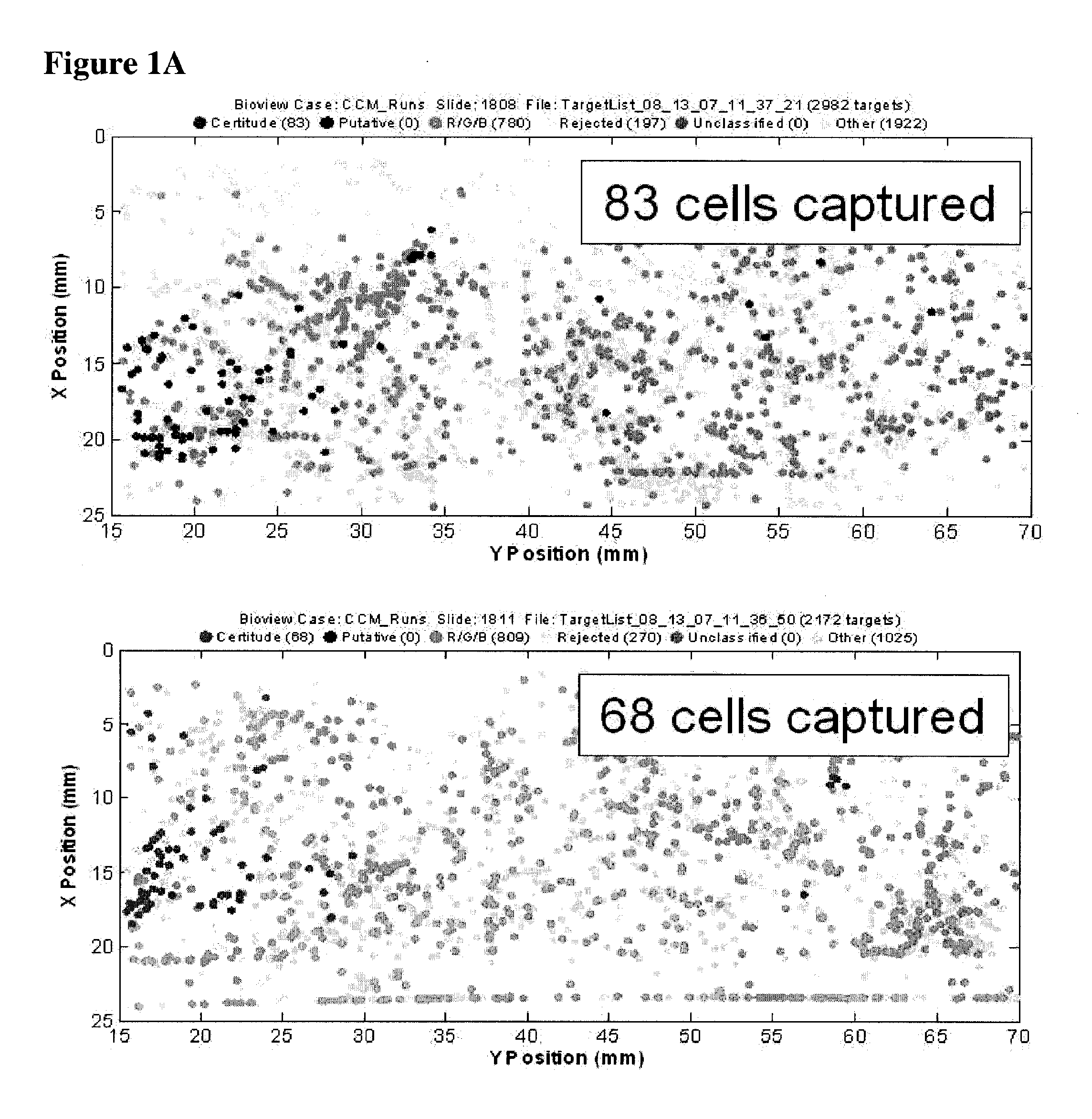
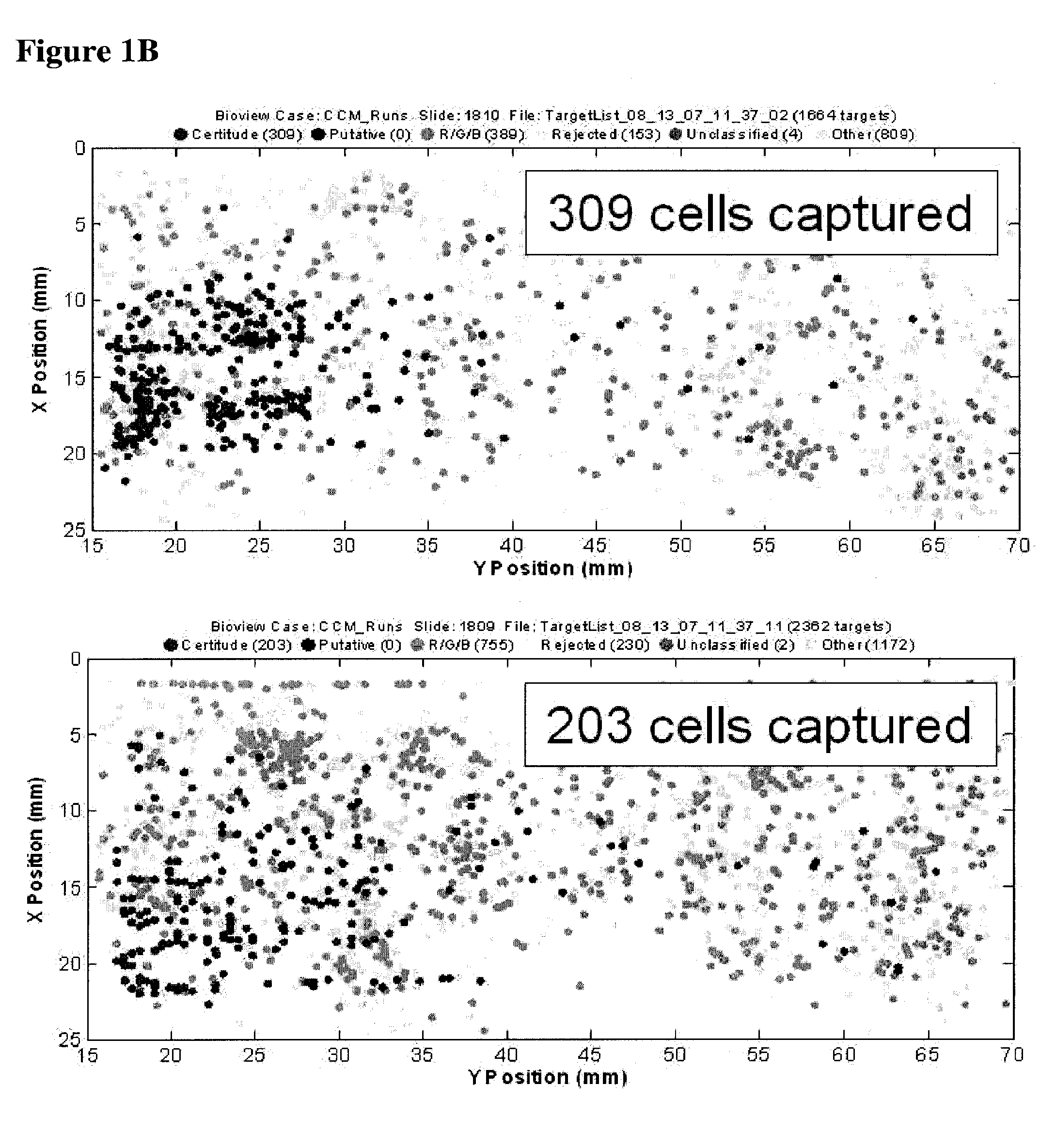
[0062]The example depicted in FIG. 1 illustrates a method and device for isolating cancer cells. The affinity-tagged ligand (ATL) in this example comprised biotin as the affinity tag. The ligand was anti-Epithelial Cell Adhesion Molecule (EpCam) antibody which recognizes an epithelial cellular marker. For the experiment, H1650 lung cancer cells were incubated with biotinylated goat anti-EpCam antibody for 1 hour on ice (FIG. 1B). Approximately 1000 cells were input into each experiment. The left and right images represent replicate experiments. As a negative control, H1650 lung cancer cells were incubated with biotinylated goat IgG antibody under the same or similar conditions (FIG. 1A). Following the incubation, the cells were washed and run through an array of obstacles [comprising a restricted gap—Chip 7, please confirm] chip coated with Neutravidin™. Cells were then fixed and visualized by fluorescence micro...

example 2

[0064]Capture of cells after solution phase binding of the cells with an affinity-tagged ligand

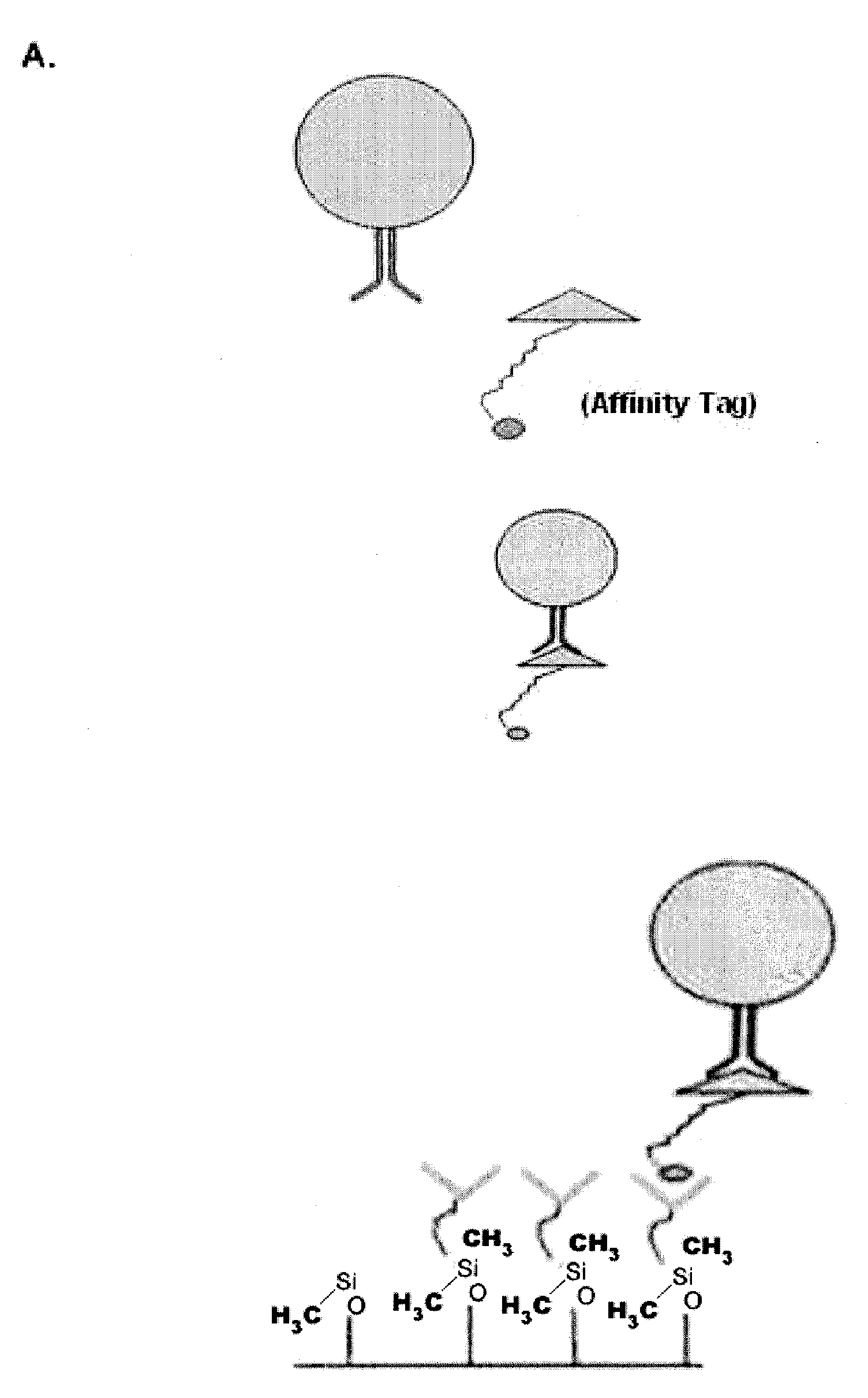
[0065]The examples depicted in FIG. 2 illustrate the capture of cells labeled with an affinity tag before being subjected to a microfluidic device. FIG. 2A is a diagram depicting a cell being contacted with an affinity-tagged ligand in solution (top panel) and the capture of such cell with a microfluidic device coated with a capture moiety capable of recognizing the affinity-tag (lower panel).

[0066]FIG. 2B illustrates the capture of cells bound to EGF peptide tagged with biotin. In this example, MDA-MB-231 cells (a breast cell line which expresses low levels of EpCam) were contacted with biotin-tagged EGF peptide in solution under conditions favorable for binding. The cells were then subjected to a chip coated with neutravidin, a capture moiety capable of recognizing the affinity tag. Following fixation, the captured cells were visualized by fluorescent microscopy. The results demonstrate ...


PUM


Abstract

Method of enriching specific cells from cellular samples are disclosed, comprising contacting in solution a cellular sample with affinity-tagged ligands (ATLs) each comprising a first ligand linked to an affinity tag, wherein the ligand selectively binds a cellular marker of the rare cells and the affinity tag can be selectively captured by a capture moiety, wherein the affinity tags do not comprise a magnetic particle; and flowing the sample through a microfluidic device comprising the capture moiety to selectively retain ATL-bound cells. Methods for enriching circulating tumor cells, and devices for enriching specific cells from cellular samples are also disclosed.

Description

BACKGROUND OF THE INVENTION[0001]Analysis of a complex mixture such as blood can be difficult but provides valuable information. For example, CD4 T cell levels can reflect the course of disease in AIDS patients, cellular lymphoid and myeloid markers can aid leukemia and lymphoma diagnoses, and the presence of errant epithelial cells in the blood may signify a metastasizing cancer.[0002]Such complex mixtures can be analyzed by fluorescent activated cell sorting (FACS), a technology that can quantitate marked analytes and also separate them out from the mixtures. Another popular method of cell separation involves magnetically labeling a target population of cells, e.g. with ferromagnetic beads, followed by sorting the labeled cells by passing them through a receptacle positioned within a magnetic field, a process also known as magnetic activated cell sorting (MACS). Despite their widespread use, FACS and MACS technologies have disadvantages, for example, they require machinery that is...

Claims


Application Information

Patent Timeline
IPC IPC(8): G01N33/574C12N11/02C12M1/00G01N33/53
CPCG01N33/574G01N33/57492Y10S435/973Y10T436/25125Y10T436/25375Y10T436/13Y10T436/101666Y10S435/971G01N33/585G01N33/54326B01L3/502761B01L2200/0647B01L2300/0861B01L2400/086G01N1/405G01N2333/705
Inventor FORSYTH, ALLYNBARNES, HELEN
Owner GPB SCI
Why Eureka
Try Eureka