
Method for servoing a source of x-rays of a digital radiography device

a digital radiography and source technology, applied in the field of medical radiography, can solve the problems of significant degradation of the signal-to-noise ratio, the inability of dose sensors to report the status of all parts of the image, and the control of the source of x-rays, so as to prevent local overexposure or underexposure.

Active Publication Date: 2009-09-03
TROPHY SAS
View PDF1 Cites 0 Cited by
  • Summary
  • Abstract
  • Description
  • Claims
  • Application Information

AI Technical Summary

Benefits of technology

[0011]One object in particular is to exactly control the radiation dose received, while preventing local overexposures or underexposures of the resulting radiographic image.
[0013]Another object is to limit the radiation dose received by the patient without impairing the quality of the radiographic images.
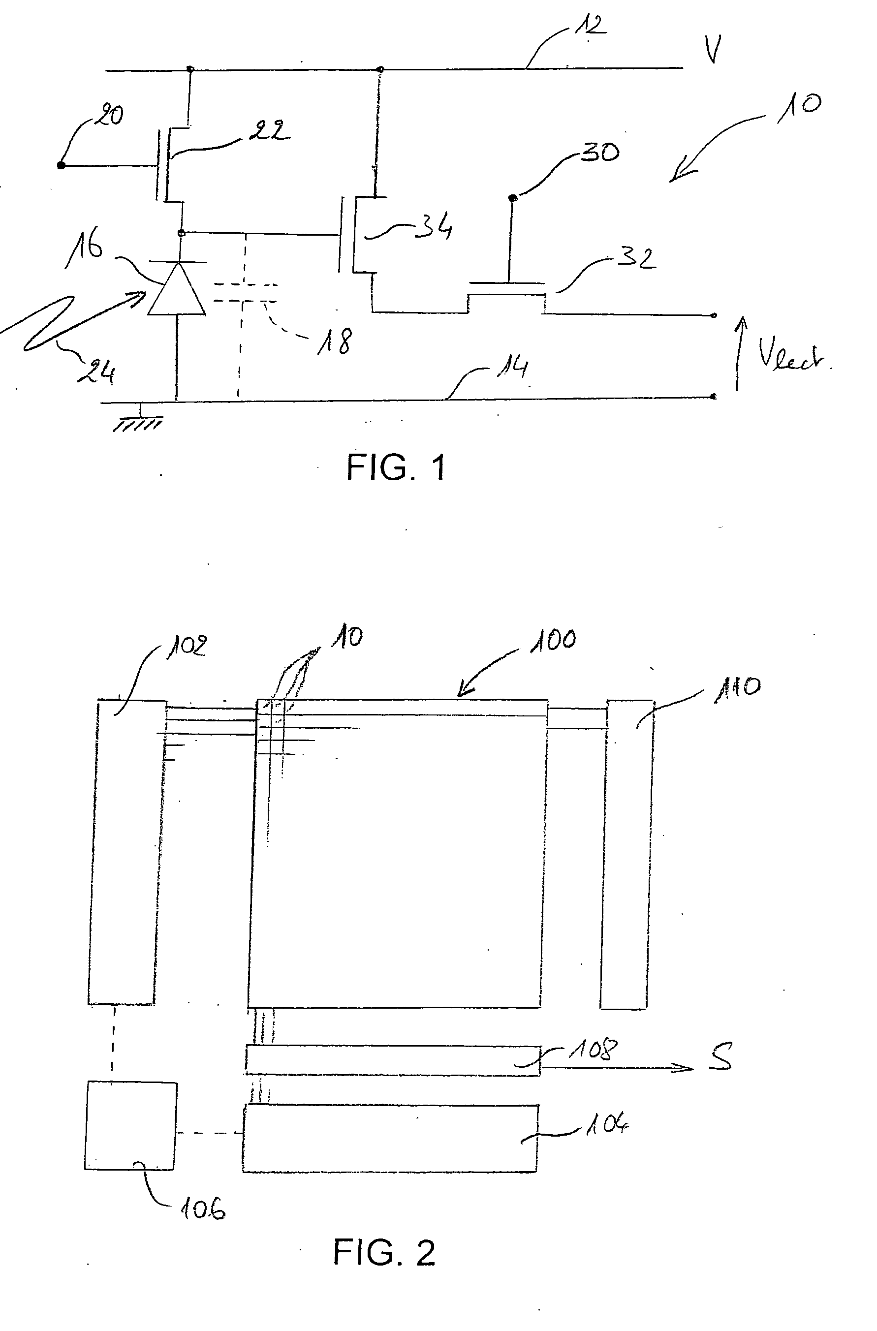
[0016]Thanks to the use of MOS-type pixels, i.e. MOS or CMOS pixels with insulated grid, and in particular pixels with separate read command, it is possible to establish and monitor the evolution of the image characteristic without disturbing pixel image integration. Indeed, refreshment is caused only when an image of sufficient quality is available, i.e. after the preset acquisition characteristic is reached.

Problems solved by technology

One of the difficulties, which exists whatever the type of sensor pixels, is to control the source of X-rays to prevent both pixel overexposure and underexposure.
However, dose sensors are incapable of reporting the status of all parts of the image, and do not prevent the existence of local over- or underexposed zones.
Inappropriate exposure in relation to the sensor's nominal exposure latitude causes either significant degradation of the signal-to-noise ratio, or pixel saturation, and impairs, in both cases, the quality of the radiographic image.

Method used


Image

  • Method for servoing a source of x-rays of a digital radiography device
  • Method for servoing a source of x-rays of a digital radiography device
  • Method for servoing a source of x-rays of a digital radiography device

Examples


Embodiment Construction

[0010]It is the object of the invention to propose a method of servoing an X-ray source and a radiography device not having the above-mentioned problems.

[0011]One object in particular is to exactly control the radiation dose received, while preventing local overexposures or underexposures of the resulting radiographic image.

[0012]Another object is to control the source to optimize the exposure for a certain tissue type.

[0013]Another object is to limit the radiation dose received by the patient without impairing the quality of the radiographic images.

[0014]To achieve these objects, the invention's aim is more precisely a method of servoing an X-ray source in a radiography device comprising a MOS-type image sensor with pixels provided with separate read and refreshment commands, respectively, in which, during a radiography operation, read commands of a plurality of image sensor pixels are called repeatedly, while maintaining an emission of X-rays, so as to establish, in response to ea...


PUM


Abstract

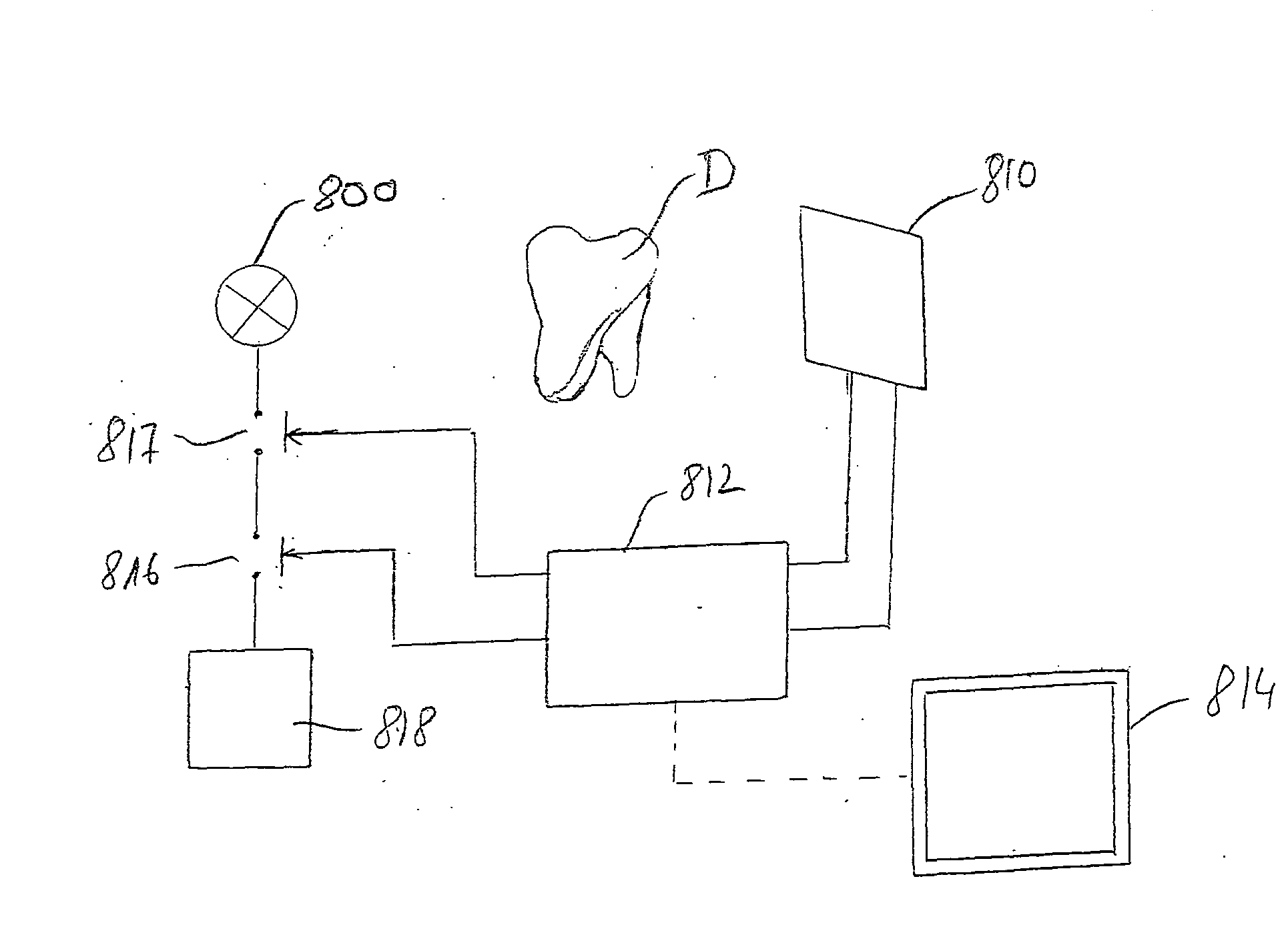
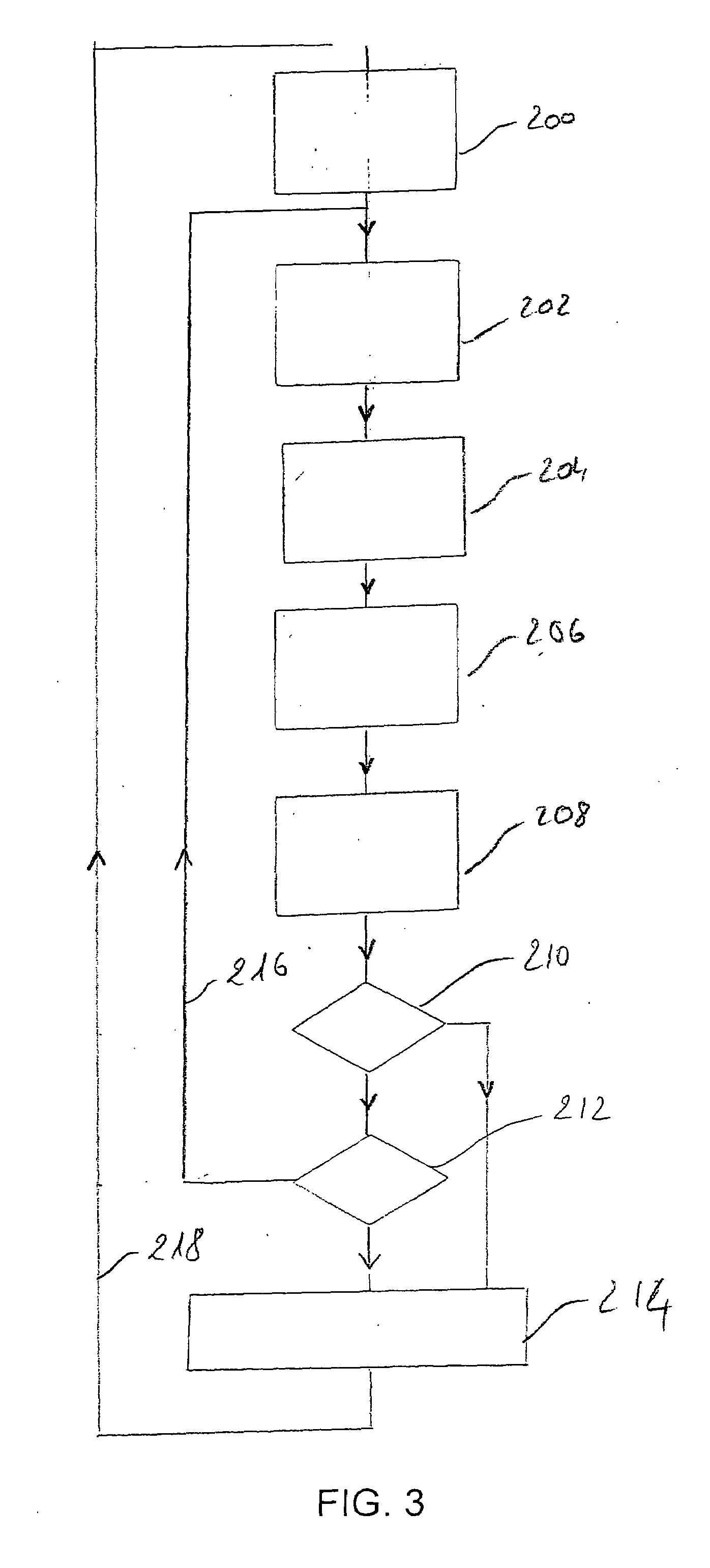
The invention relates to a radiographic device and method of servoing an X-ray source (800) in a radiography device comprising a MOS-type image sensor (810) with pixels provided with separate read and refreshment commands, respectively, in which, during a radiography operation, read commands of a plurality of image sensor pixels are called repeatedly, while maintaining a source of X-rays power-supplied, so as to establish, in response to each reading, at least one image acquisition characteristic, and in which the emission of X-rays is interrupted when the image acquisition characteristic corresponds to a preset image acquisition characteristic.

Description

TECHNICAL FIELD[0001]The present invention relates to a method for servoing a source of X-rays of a digital radiography device and digital radiography equipment using a servoed source. The invention has applications in the field of medical radiography, and in particular that of dental radiography.STATE OF THE PRIOR ART[0002]Digital radiography devices have a sensor capable of generating electrical signals that are a function of the radiation intensity, and more precisely the energy received at any point on a sensitive surface. Therefore, the sensor's sensitive surface is subdivided into a matrix of sensitive elements that, by analogy with the image capable of being formed based on the sensor's signal, are also called “pixels”. The sensitive surface is linked to a scintillator that converts the X-rays received by the sensor into radiation whose wavelength is compatible with the pixels' sensitivity spectrum.[0003]Two separate technologies are used to constitute the sensor's pixels. Th...

Claims


Application Information

Patent Timeline
IPC IPC(8): A61B6/14H05G1/64
CPCA61B6/032A61B6/14A61B6/4233H04N5/3745H05G1/44H04N5/32A61B6/482H04N25/77A61B6/51H04N25/30A61B6/00
Inventor INGLESE, JEAN-MARCBOTHOREL, SYLVIE MARIE GISELELEMAIRE, ALAIN PAULBOUCLY, ALAIN
Owner TROPHY SAS
Learn More