Visualization of coronary vein procedure

a technology for visualizing procedures and coronary veins, applied in balloon catheters, surgery, other medical devices, etc., can solve problems such as significant health risks of contrast media, renal dysfunction or failure,

Inactive Publication Date: 2009-09-17
OSPREY MEDICAL
View PDF5 Cites 64 Cited by
  • Summary
  • Abstract
  • Description
  • Claims
  • Application Information

AI Technical Summary

Problems solved by technology

Contrast media may have significant health risks if permitted to flow systemically to the patient's org

Method used


Image

  • Visualization of coronary vein procedure
  • Visualization of coronary vein procedure
  • Visualization of coronary vein procedure

Examples


Embodiment Construction

[0028]Referring now to the several drawing figures in which identical elements are numbered identically throughout, a description of a preferred embodiment of the present invention will now be provided.

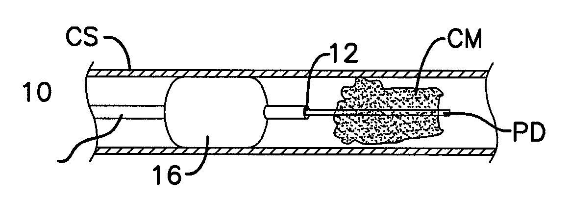
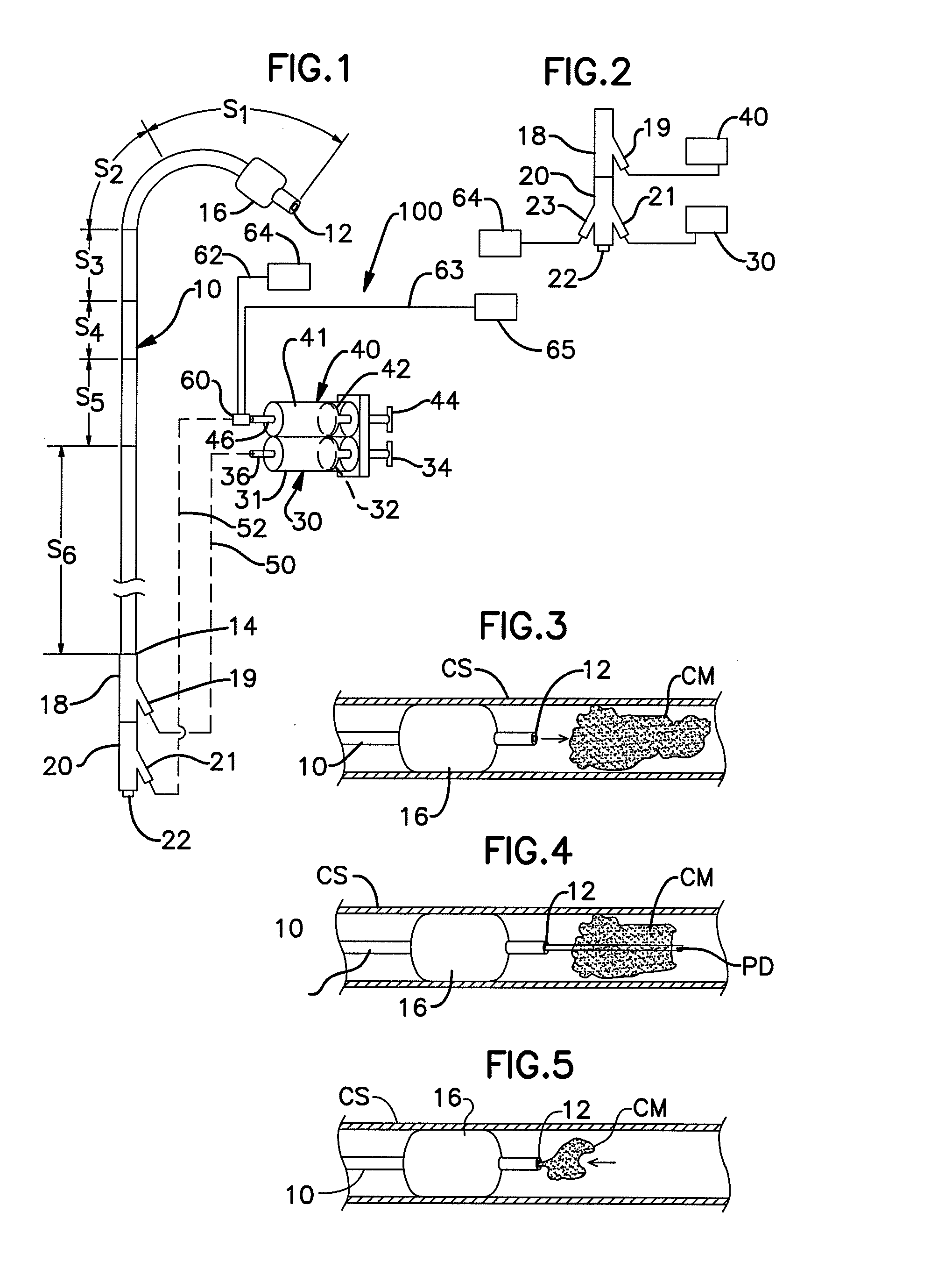
[0029]FIG. 1 shows a system 100 including a catheter 10 according to the present invention. In the preferred embodiment, the catheter 10 is used to inject a contrast media into a coronary sinus or other coronary vein. Following such injection, the catheter may be used for advancing medical procedure tools through a lumen of the catheter 10. Such procedures may include lead placement for bi-ventricular pacing or measuring devices for mitral valve re-shaping, by way of non-exhaustive examples. Following such procedure, the catheter 10 is used to remove the injected contrast media from the coronary sinus.

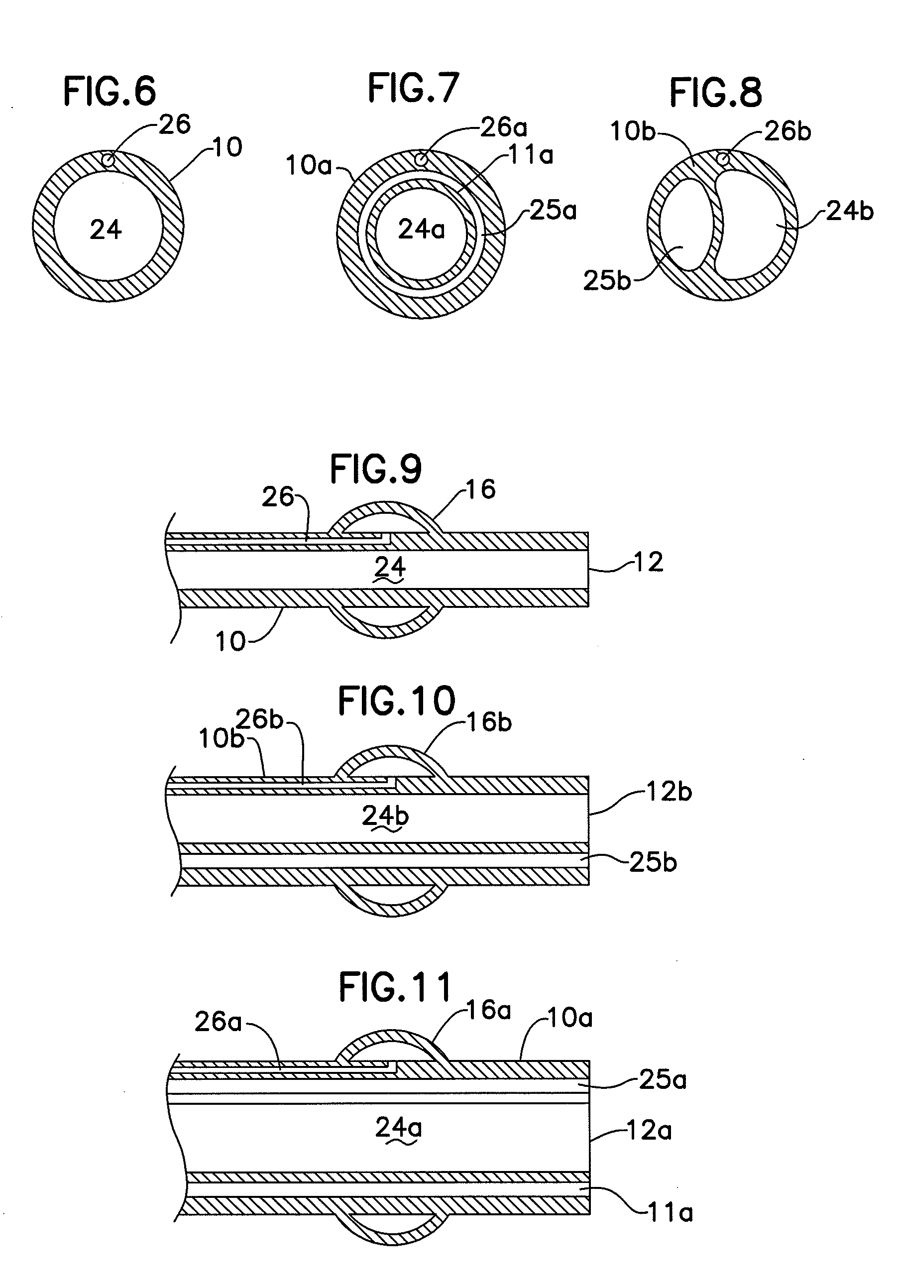
[0030]The catheter 10 is a long flexible and hollow tubular member 10 which has an opening at the distal end 12 and proximal end 14. The catheter 10 may be sized for delivery through a j...


PUM


Abstract

Visualization of a medical procedure to be performed in a coronary vessel (such as a coronary sinus) of a patient is achieved by the steps of: identifying a coronary vessel for a procedure and placing a catheter within the coronary vessel. The coronary vessel is occluded at a site proximal to a distal end of the catheter. A contrast medium is injected into the coronary vessel through the catheter distal end for visualization of the procedure within the coronary vessel. Following the procedure, the contrast medium is removed through the catheter distal end and occlusion of the coronary vessel is discontinued.

Description

I. BACKGROUND OF THE INVENTION[0001]1. Field of the Invention[0002]This invention pertains method and apparatus for visualizing procedures performed in a coronary vessel such as a coronary vein.[0003]2. Description of the Prior Art[0004]Angiography procedures are widely used for assessing patency of coronary arteries. These procedures may also be used for other purposes—for example, in stent placement or other procedures. In such a procedure, a contrast medium is injected into a coronary artery proximal to a suspected site of a coronary obstruction.[0005]Procedures are also developed (or in development) for performing procedures in coronary veins such as the coronary sinus. By way of non-limiting example, such procedures include lead placement for bi-ventricular pacing and procedures such as those shown in U.S. Pat. No. 6,964,683 to Kowalsky et al. issued Nov. 15, 2005 and U.S. Pat. No. 6,966,926 to Mathis issued Nov. 22, 2005 for re-shaping a mitral valve.[0006]As in coronary arte...

Claims


Application Information

Patent Timeline
IPC IPC(8): A61M31/00A61M25/10A61F2/958
CPCA61B17/1204A61B17/12109A61B17/12136A61M1/0023A61M2025/1052A61M5/204A61M25/0668A61M25/1011A61M2005/3128A61M5/007
Inventor SHAPLAND, JAMES EDWARDDOAN, TUAN MINHALFERNESS, CLIFTON A.
Owner OSPREY MEDICAL
Why Eureka
Try Eureka