
System for Generation of a Composite Medical Image of Vessel Structure

a medical image and composite technology, applied in image enhancement, image analysis, instruments, etc., can solve problems such as unwelcome mis-registration artifacts, and achieve the effect of reducing artifacts

Active Publication Date: 2009-10-15
PIXART IMAGING INC
View PDF3 Cites 50 Cited by
  • Summary
  • Abstract
  • Description
  • Claims
  • Application Information

AI Technical Summary

Benefits of technology

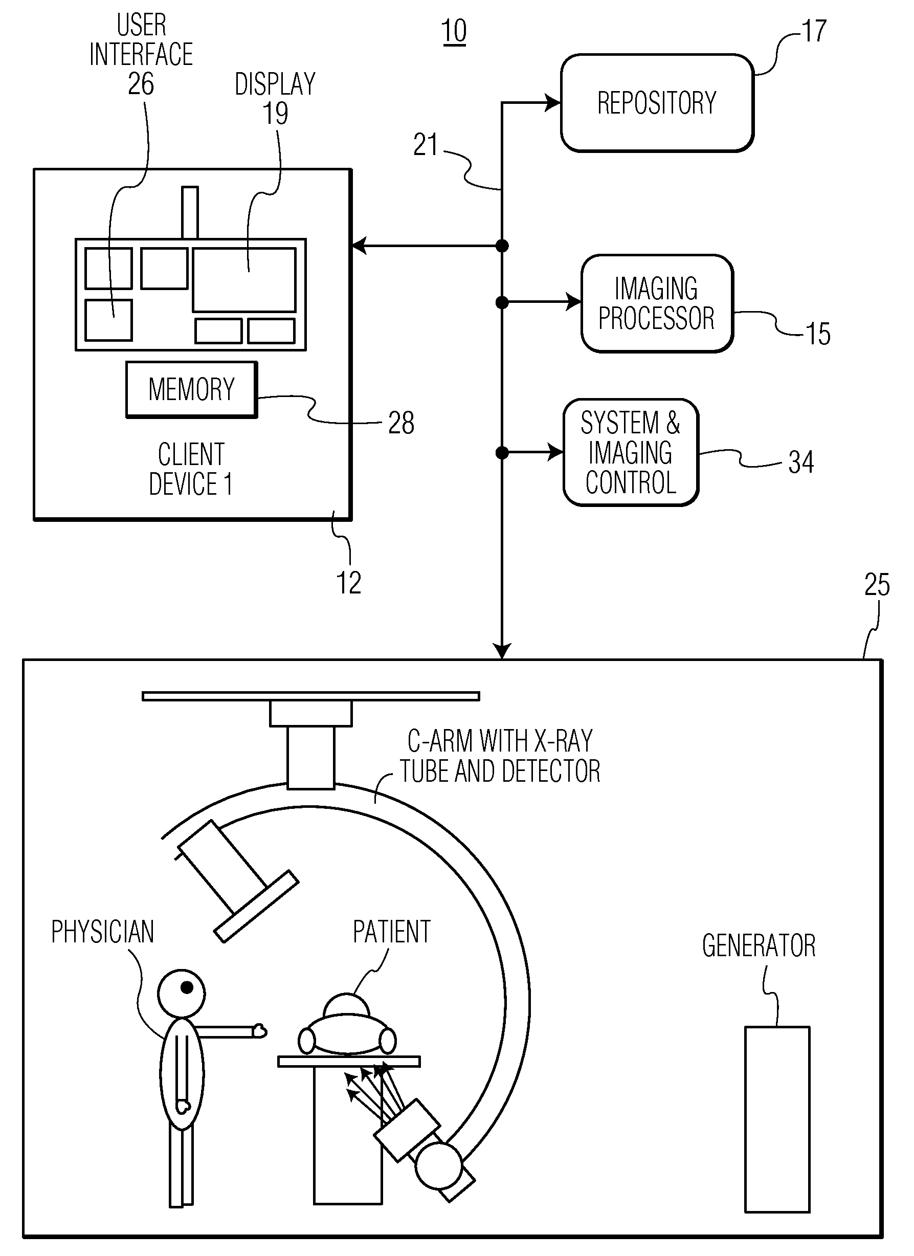
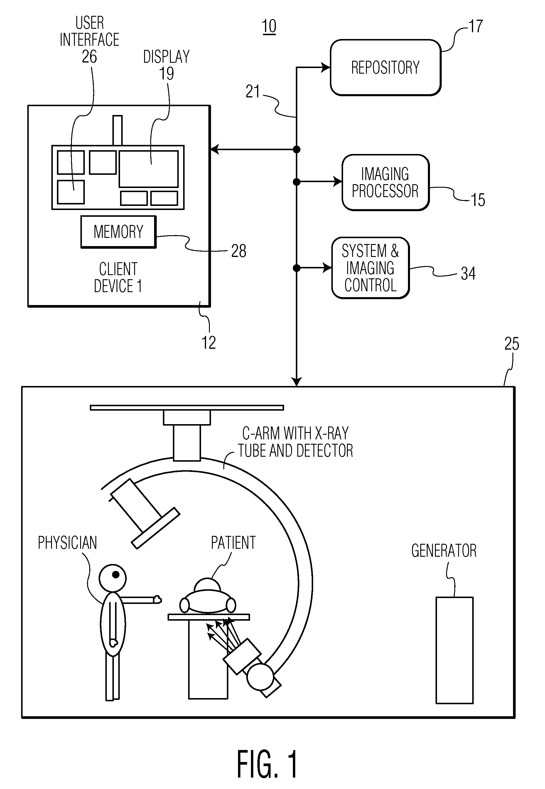
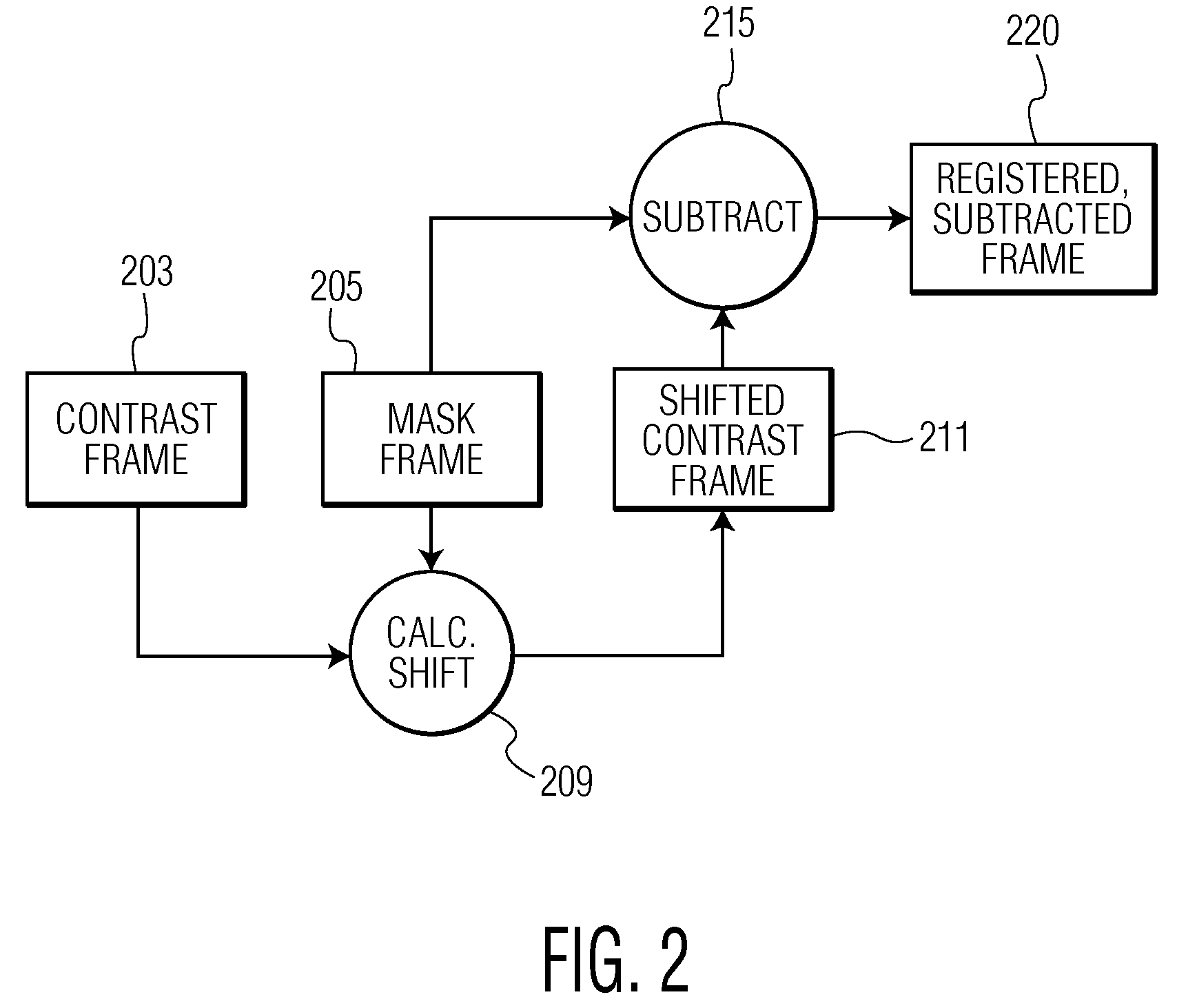
[0004]A system reduces artifacts introduced by patient or table motion during generation of a composite image visualizing contrast agent flow over a subset of image frames acquired during an angiography imaging study that represent a specific time period within a period of imaging acquisition. A system for generation of a composite medical image of vessel structure, includes an imaging device for acquiring multiple sequential images of vessel structure of a portion of patient anatomy in the presence of a contrast agent. An imaging processor aligns individual images of the multiple sequential images with a single particular mask image containing background detail of the portion of patient anatomy in the absence of contrast agent. The imaging processor forms data representing multiple digitally subtracted images by subtracting data representing the single particular mask image from aligned individual images of the multiple sequential images. The imaging processor associates different individual images of the multiple digitally subtracted images with different corresponding visual attributes (e.g. colors, hues) and combines data representing the digitally subtracted images to form a composite image. A reproduction device presents the composite image to a user.

Problems solved by technology

Therefore, image related motion that occurs due to, slight patient movement or slight X-ray table motion, for example, causes undesired mis-registration artifacts to appear in a resulting blood flow representative composite image.

Method used


Image

  • System for Generation of a Composite Medical Image of Vessel Structure
  • System for Generation of a Composite Medical Image of Vessel Structure
  • System for Generation of a Composite Medical Image of Vessel Structure

Examples


Embodiment Construction

[0010]A system reduces artifacts introduced by patient movement or motion of an X-ray unit patient support table during generation of an image visualizing contrast agent flow from a subset of digitally subtracted image frames comprising images acquired during an angiography imaging study. Individual images of multiple sequential images acquired during a specific time period within the duration of acquisition of the angiography imaging study are aligned with a single particular mask image that contains background detail of a portion of patient anatomy in the absence of contrast agent. The particular mask image is subtracted from the individual images of the multiple sequential images. Different individual images of the resultant multiple digitally subtracted images are associated with different corresponding visual attributes (e.g. colors, hues) and combined to form a composite image.

[0011]A processor as used herein is a device for executing stored machine-readable instructions for p...


PUM


Abstract

A system reduces artifacts introduced by patient or table motion during generation of a composite image visualizing contrast agent flow. A system for generation of a composite medical image of vessel structure, includes an imaging device for acquiring multiple sequential images of vessel structure of a portion of patient anatomy in the presence of a contrast agent. An imaging processor aligns individual images of the multiple sequential images with a single particular mask image containing background detail of the portion of patient anatomy in the absence of contrast agent. The imaging processor forms data representing multiple digitally subtracted images by subtracting data representing the single particular mask image from aligned individual images of the multiple sequential images. The imaging processor associates different individual images of the multiple digitally subtracted images with different corresponding visual attributes and combines data representing the digitally subtracted images to form a composite image. A reproduction device presents the composite image to a user.

Description

[0001]This is a non-provisional application of provisional application Ser. No. 61 / 044,114 filed Apr. 11, 2008, by J. Baumgart.FIELD OF THE INVENTION[0002]This invention concerns a system for generation of a composite medical image of vessel structure and contrast agent flow (representing blood flow) by allocating different visual attributes (e.g., different colors) to individual digitally subtracted images formed by subtracting background detail and by combining the individual digitally subtracted images to form a single composite image.BACKGROUND OF THE INVENTION[0003]In order to study contrast agent flow, which is representative of blood flow, through vessels in an angiography image sequence, it is useful to generate a static representation of flow using a color spectrum to represent the time at which contrast agent reached a certain state within vessels (e.g., first entered, reached a peak, no longer seen). For angiography, a typical color spectrum uses red to represent early bl...

Claims


Application Information

Patent Timeline
IPC IPC(8): G06K9/00
CPCG06T5/50G06T2207/20224G06T2207/30104G06T2207/10121
Inventor BAUMGART, JOHN
Owner PIXART IMAGING INC
Learn More