
X-ray imaging with X-ray markers that provide adjunct information but preserve image quality

a technology of adjunct information and x-ray markers, applied in the field of x-ray imaging, can solve the problems of process typically having a relatively high error rate, breast cancer and other breast lesions remain a significant threat to women's health, and the process is typically not accura

Active Publication Date: 2009-10-29
HOLOGIC INC
View PDF36 Cites 161 Cited by
  • Summary
  • Abstract
  • Description
  • Claims
  • Application Information

AI Technical Summary

Benefits of technology

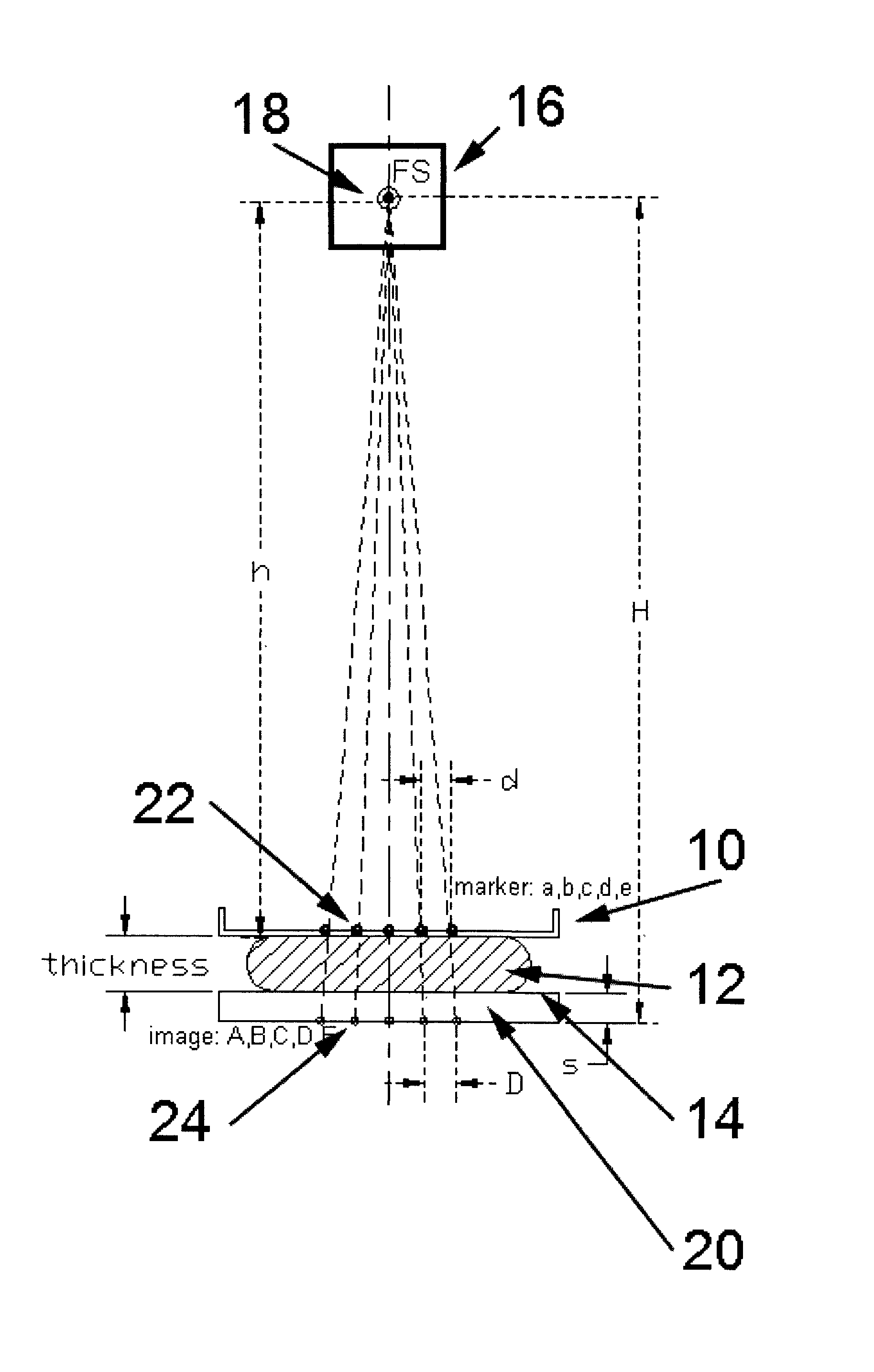
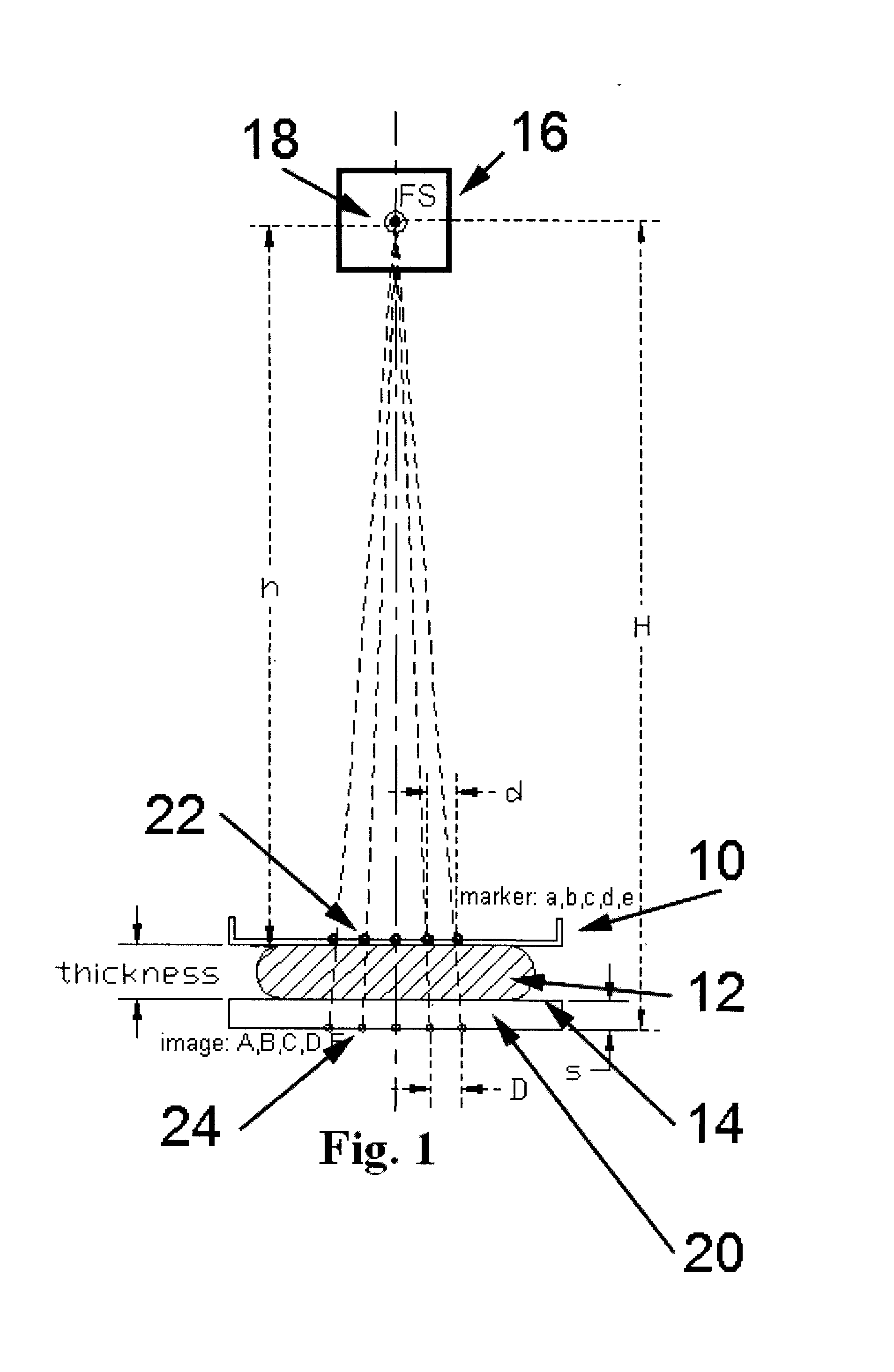
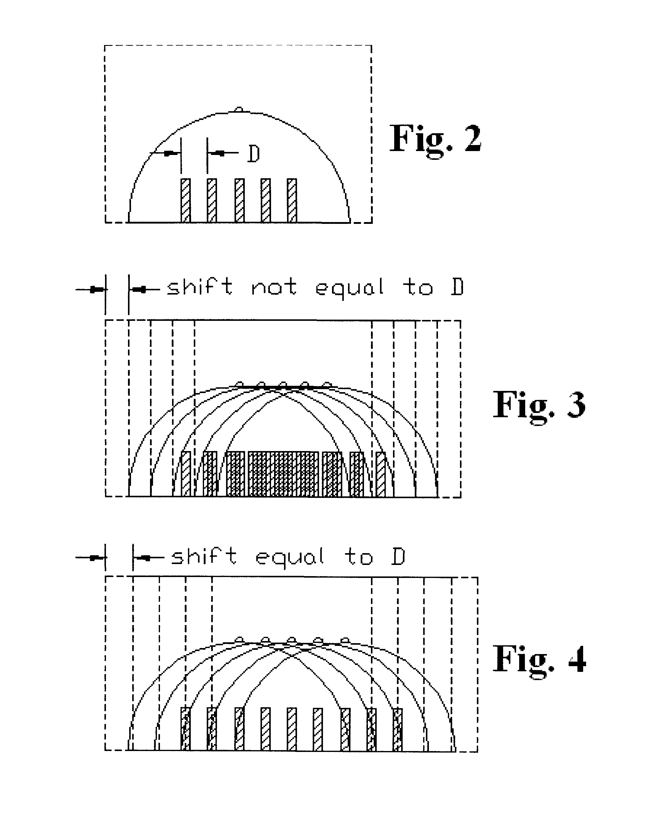
[0006]In one non-limiting example, a breast imaging method comprises x-ray imaging a pattern of markers on a compression paddle and a patient's breast immobilized between the paddle and an x-ray imaging receptor to form a composite x-ray image in which the x-ray image of markers is composited with the x-ray image of the breast, computer-processing the composite x-ray image to derive geometric information regarding the imaged markers, computer-processing the geometric information to derive thickness information related to a thickness of the immobilized breast or other information related to the position of the pattern of markers in space or relative to system components, and using the thickness or spatial position information for improving breast imaging. The imaged markers may be invisible within the breast outline when viewing the composite image in typical clinical settings or, in alternative embodiments, they may be visible in the composite image. The method can further include removing some or essentially all the contribution of the markers from the composite x-ray image.
[0007]As another non-limiting example, an apparatus comprises a compression paddle with a pattern of markers that immobilizes a patient's breast against a breast platform, an x-ray source on one side of the paddle and platform and an x-ray imaging receptor on the other side, a processor receiving image information from the receptor related to a composite x-ray image of the markers and breast in which the markers are not visible in a clinical setting, said processor computer-processing the composite image to derive geometric information regarding the markers therein and computer-processing the geometric information to derive thickness information related to thickness of the immobilized breast, and said processor using the thickness information to improve imaging the breast. The processor can further process the composite image to completely or essentially remove some or essentially all contribution of the imaged markers.
[0009]In addition to or instead of being on the compression paddle, similar markers can be placed on other objects. For example, markers can be placed on the magnification table that is placed between the normal breast platform and the breast to increase the distance between the breast and the imaging plane and thus magnify the breast image. Markers can be placed on the normal breast platform if it desirable to calculate or confirm the spacing between the breast platform and the image plane. In general, markers can be placed on any object that is between the x-ray tube and the image plane of the x-ray receptor to help calculate the distance between the object and the image plane or other objects.

Problems solved by technology

Breast cancer and other breast lesions continue to be a significant threat to women's health.
It has been proposed to use encoders to measure the geometric height of a breast compression paddle and use the result to estimate the geometric thickness of the breast but this process typically has a relatively high error because of factors such as tilting and geometric distortions of the paddle as it compresses the breast, and because the encoders must be calibrated.

Method used


Image

  • X-ray imaging with X-ray markers that provide adjunct information but preserve image quality
  • X-ray imaging with X-ray markers that provide adjunct information but preserve image quality
  • X-ray imaging with X-ray markers that provide adjunct information but preserve image quality

Examples


Embodiment Construction

[0030]This patent specification describes methods and systems in which the geometric thickness of a body part such as the breast that is being x-rayed is accurately determined and used to improve imaging in a manner that does not inconvenience the patient or the health professional. It also describes similar processes to determine other thicknesses, heights, or distances between objects in x-ray examination procedures.

[0031]In describing preferred embodiments illustrated in the drawings, specific terminology is employed for the sake of clarity. However, this patent specification is not intended to be limited to the specific terminology so selected and it is to be understood that each specific element includes all technical equivalents that operate in a similar manner. In addition, a detailed description of known functions and configurations will be omitted when it may obscure the subject matter of the invention described in the appended claims.

[0032]As illustrated in the example of ...


PUM


Abstract

A method and an apparatus for estimating a geometric thickness of a breast in mammography / tomosynthesis or in other x-ray procedures, by imaging markers that are in the path of x-rays passing through the imaged object. The markings can be selected to be visible or to be invisible when the composite markings / breast image is viewed in clinical settings. If desired, the contribution of the markers to the image can be removed through further processing. The resulting information can be used determining the geometric thickness of the body being x-rayed and thus setting imaging parameters that are thickness-related, and for other purposes. The method and apparatus also have application in other types of x-ray imaging.

Description

TECHNICAL FIELD[0001]This patent specification relates to x-ray imaging and more specifically to accurately finding the geometric thickness of an object being x-rayed through including markers in the x-ray path and computer-processing the composite markers / object x-ray image to identify the markers and to use the resulting information to calculate geometric object thickness and improve imaging.BACKGROUND[0002]Breast cancer and other breast lesions continue to be a significant threat to women's health. X-ray mammography currently is the most widely used tool for early detection and diagnosis, and is the modality approved by the U.S. Food and Drug Administration to screen for breast cancer in women who do not show symptoms of breast disease. A typical x-ray mammography system compresses and immobilizes a patient's breast on a breast platform positioned between an x-ray source and an x-ray imager, and takes a projection x-ray image (called here a conventional mammogram or simply mammog...

Claims


Application Information

Patent Timeline
IPC IPC(8): A61B6/04
CPCA61B5/1075A61B6/0414A61B6/583A61B6/544A61B6/502
Inventor REN, BAORUISMITH, ANDREW P.JING, ZHENXUESTEIN, JAYDEFREITAS, KENNETH F.
Owner HOLOGIC INC
Learn More