

Patient support table for use in magnetic resonace imaging
- Summary
- Abstract
- Description
- Claims
- Application Information
AI Technical Summary
Benefits of technology
Problems solved by technology
Method used
Image

Examples
Embodiment Construction
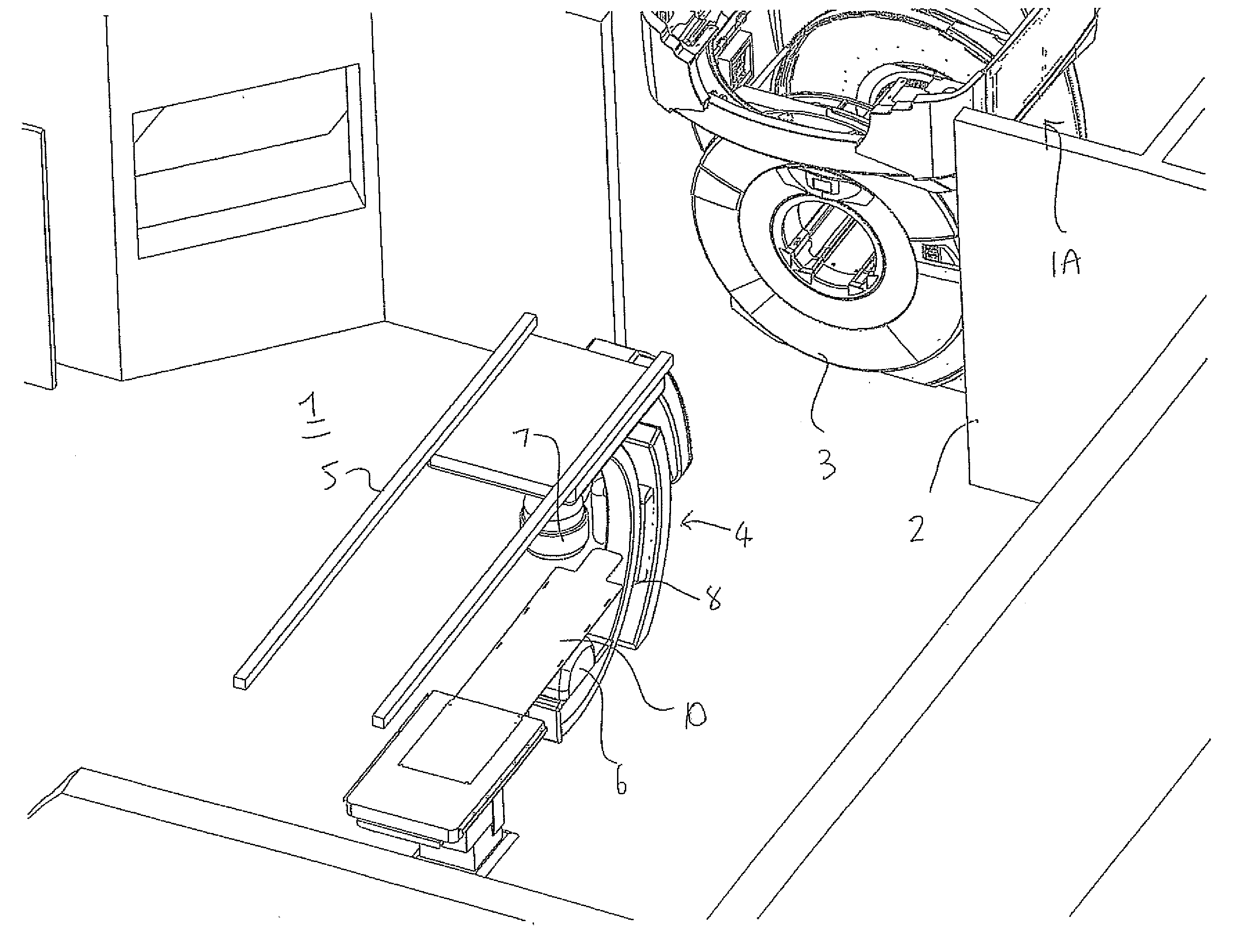
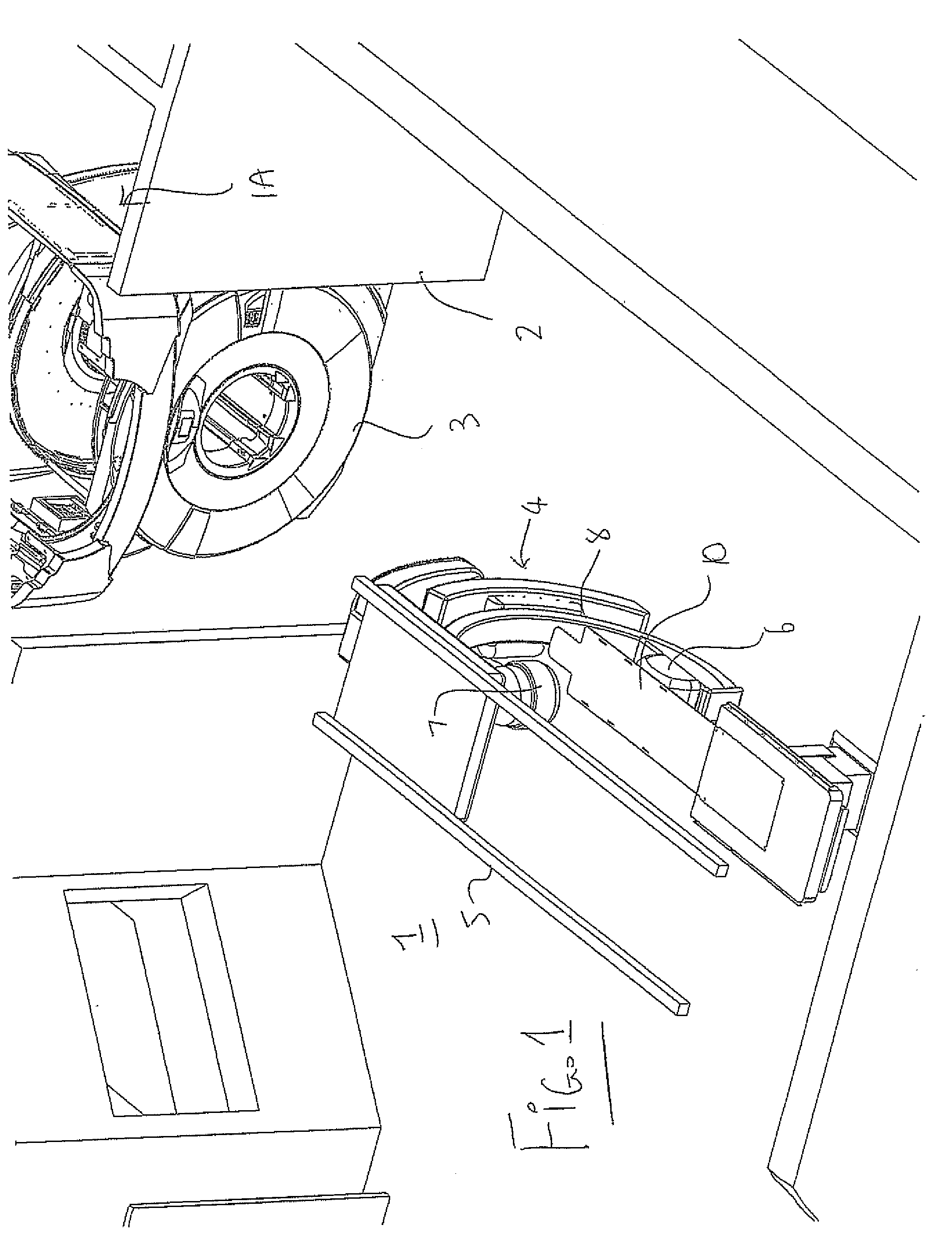
[0109]Reference may be made to the abovementioned Published PCT Application WO07147233A1 of the present Applicants published Dec. 27, 2007 and entitled ROTATABLE INTEGRATED SCANNER FOR DIAGNOSTIC AND SURGICAL IMAGING APPLICATIONS in which is disclosed details of the construction of an MRI magnet suitable for use in the present arrangement. The disclosure of this document is incorporated herein by reference.
[0110]In FIG. 1 is shown an arrangement for carrying out Magnetic Resonance Imaging and X-ray imaging of a patient while the patient remains stationary on a patient support table. The arrangement provides a room 1 in which is mounted a patient support table 10 with doors 2 at one side of the room for entry into the room of the magnet 3 of an MR imaging system from a magnet bay 1A. The room contains an X-ray imaging system 4 mounted on rails 5 and includes an X-ray transmitter 6 and receiver 7 mounted on a C-shaped support 8. The X-ray system is of a conventional construction comme...
PUM
 Abstract
Description
Claims
Application Information