Method and system for breast cancer screening

a breast cancer and screening method technology, applied in the field of breast cancer screening, can solve the problems of patients exposed to ionizing radiation, uncomfortable procedures, and face disadvantages, and achieve the effect of reliable and improved screening tools

Inactive Publication Date: 2010-01-21
TOMTEC IMAGING SYST
View PDF4 Cites 28 Cited by
  • Summary
  • Abstract
  • Description
  • Claims
  • Application Information

AI Technical Summary

Benefits of technology

[0011]It is an object of the present invention to provide a method and a system for breast cancer screeni

Problems solved by technology

Mammography is the standard screening method for breast cancer; however, it faces disadvantages especially in detecting calcifications in young patients.
The disadvantages of mammography are that the patients are exposed to ionizing radiation and that the procedure is uncomfortable due to breast compression.
Additionally, there are limitations in detecting calcifications in dense breasts.
So far, breast ultrasound has been established as a valuable diagnostic test, but not as a reliable screening method because it is highly operator dependent and thus cannot be standardized easily.
However, up to now microcalcifications can be made visible by ultrasound for breast screening only very poorly since the ultrasound resolution has been limited to about 1 millimeter.

Method used


Image

  • Method and system for breast cancer screening
  • Method and system for breast cancer screening
  • Method and system for breast cancer screening

Examples


Embodiment Construction

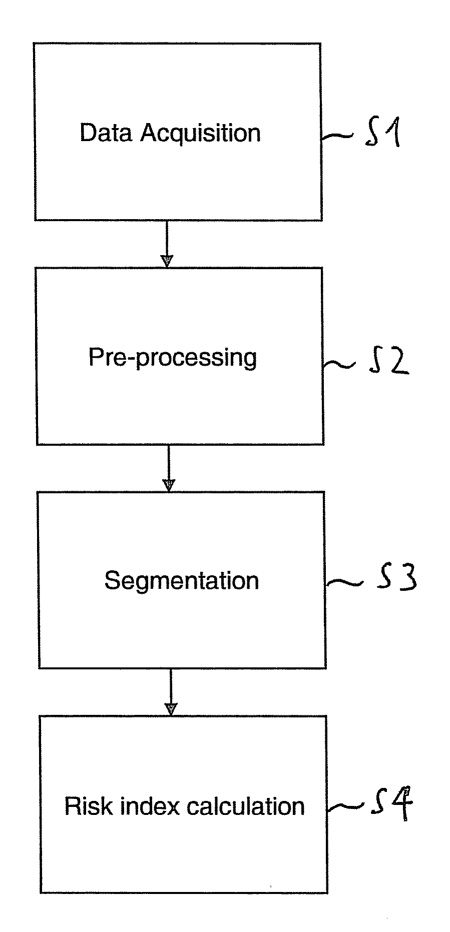
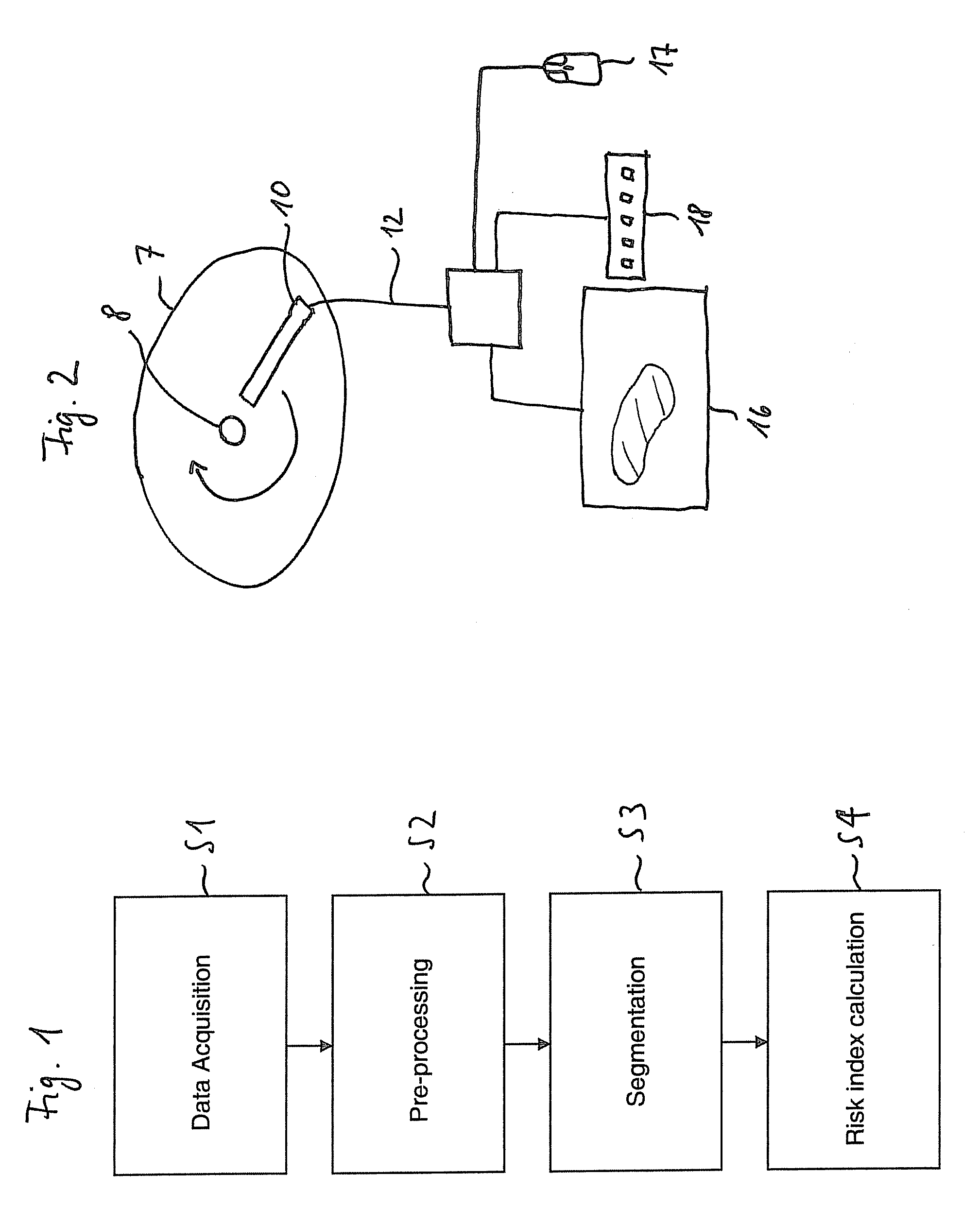
[0053]According to FIG. 1, in step S1 of a first, preferred embodiment of the method according to the invention, data acquisition is performed. The patient, i.e. the woman to be scanned, is placed e.g. in a supine, prone or sedentary position for an optimal scanning of the breast, in particular of the gland tissue, such as the lobes, the lobules, and the milk ducts. As depicted in FIG. 2, an ultrasonic transducer 10 which has an elongate shape is placed on the breast 7 of the woman such that one end of the transducer 10 is disposed close to the nipple 8 of the breast 7 and the other end thereof is disposed distant therefrom, the transducer 10 being oriented radially on the breast. The transducer 10 is preferably rotated by 360°, and during the whole rotation ultrasound waves are emitted and the echoes from the woman are recorded by the transducer 10. Alternatively, the transducer 10 may be rotated by an angle of less than 360°, e.g. only by 90° for scanning one quadrant. The radial ...


PUM


Abstract

The invention relates to a system for breast cancer screening and a corresponding method carried out with the system, the method comprising the following steps:
    • a) providing an image of a predetermined region of a breast of a woman,
    • b) determining the glandular volume in the scanning image, and
    • c) calculating the absolute glandular tissue amount of the breast from the glandular volume,
    • wherein the absolute glandular tissue amount can be used as a risk index which provides an indication about the risk of the woman of having breast cancer.

Description

TECHNICAL BACKGROUND[0001]The present invention relates to a method and a system for breast cancer screening, in particular for breast cancer screening by ultrasonography or magnetic resonance imaging (MRI). In general, the method and the system of the invention are applicable to any kind of data obtained with various imaging modalities.[0002]In order to further reduce medical treatment and mortality due to breast cancer, it is highly desirable to achieve a better early diagnosis thereof, which may be accomplished inter alia by better screening tools.[0003]Mammography is the standard screening method for breast cancer; however, it faces disadvantages especially in detecting calcifications in young patients. The cost-effective, easy-to-use and non-ionizing modality ultrasound can be used to resolve different tissues within the breast such as fat or glandular tissue. Glandular tissue, or briefly termed gland tissue, is in particular tissue such as the lobes, the lobules, and the milk ...

Claims


Application Information

Patent Timeline
IPC IPC(8): G06K9/00
CPCG06T7/0012G06T2207/30068G06T2207/10132G06T2207/10088
Inventor BIRNHOLZ, JASONGALUSCHKY, CHRISTIANSCHRECKENBERG, MARCUSMUMM, BERNHARD
Owner TOMTEC IMAGING SYST
Why Eureka
Try Eureka