Method for rapid detection of lymphatic filariasis
a lymphatic filariasis and lymphatic system technology, applied in the field of rapid detection of lymphatic filariasis, can solve the problems of inability to detect cryptic infections, inability to detect many positive cases, and severe lack of sensitivity of traditional methods, and achieve the effect of simple and rapid diagnosis
- Summary
- Abstract
- Description
- Claims
- Application Information
AI Technical Summary
Benefits of technology
Problems solved by technology
Method used
Image
Examples
first embodiment
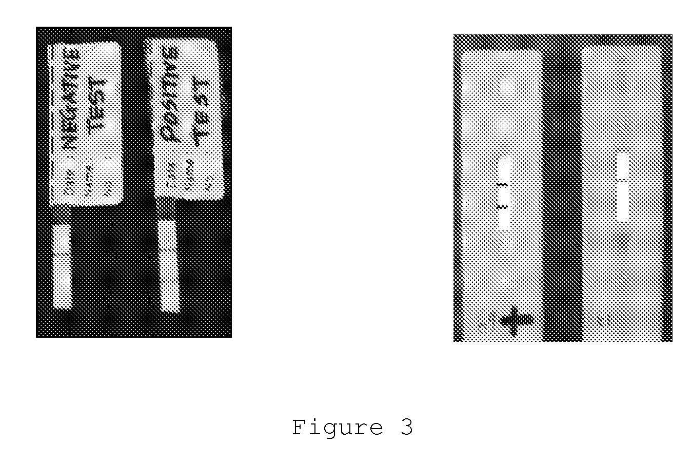
[0053]In the present invention, a buffer is added to reconstitute dried mouse monoclonal anti-human IgG4 antibody conjugated to colloidal gold in a microwell. Then, a biological sample such as blood, serum, plasma, urine or tears is introduced to the sample receiving end of an absorbent nitrocellulose membrane and is allowed to migrate laterally via capillary action towards the reaction zone of the membrane. The anti-filarial IgG4 antibodies present in the sample will bind to the SXP / SXP-1 recombinant antigen immobilized within the reaction zone, forming an antibody-antigen complex or immunocomplex. Next, the absorbent nitrocellulose membrane is placed in the microwell containing the reconstituted mouse monoclonal anti-human IgG4 antibody conjugated to colloidal gold. The mouse monoclonal anti-human IgG4 antibody conjugated to colloidal gold absorbs through the membrane and migrates to the reaction zone and binds to the antibody-antigen complex formed earlier thus forming a complex ...
second embodiment
[0054]In the present invention, a biological sample such as blood, serum, plasma, urine or tears is first introduced to the sample receiving end of the absorbent nitrocellulose membrane and is allowed to migrate laterally via capillary action towards the reaction zone of the membrane. The anti-filarial IgG4 antibodies present in the sample will bind to the SXP / SXP-1 recombinant antigen immobilized within the reaction zone, forming an antibody-antigen complex or immunocomplex. A buffer is then introduced to the reagent releasing end to reconstitute dried mouse monoclonal anti-human IgG4 antibody conjugated to colloidal gold incorporated therein. The mouse monoclonal anti-human IgG4 antibody conjugated to colloidal gold migrates to the reaction zone and binds to the antibody-antigen complex formed earlier thus forming a complex which comprises SXP / SXP-1 recombinant antigen, anti-filarial IgG4 antibodies and mouse monoclonal anti-human IgG4 antibody conjugated to colloidal gold. The pr...
third embodiment
[0055]In the present invention a biological sample such as blood, serum, plasma, urine or tears is first introduced to the sample receiving end of the absorbent nitrocellulose membrane and is allowed to migrate laterally via capillary action towards the reaction zone of the membrane. The anti-filarial IgG4 antibodies present in the sample will bind to the SXP / SXP-1 recombinant antigen immobilized within the reaction zone, forming an antibody-antigen complex or immunocomplex. A buffer is then introduced to the reagent releasing end to reconstitute dried mouse monoclonal anti-human IgG4 antibody conjugated to colloidal gold incorporated therein. The mouse monoclonal anti-human IgG4 antibody conjugated to colloidal gold migrates to the control zone and bind with the anti-mouse IgG antibody, forming a red-purplish line in the control zone. The unbound mouse monoclonal anti-human IgG4 antibody conjugated to colloidal gold will further migrate to the reaction zone and binds to the antibod...
PUM
| Property | Measurement | Unit |
|---|---|---|
| molecular weight | aaaaa | aaaaa |
| volume | aaaaa | aaaaa |
| concentration | aaaaa | aaaaa |
Abstract
Description
Claims
Application Information
 - R&D
- Intellectual Property
- Life Sciences
- Materials
- Tech Scout
- Unparalleled Data Quality
- Higher Quality Content
- 60% Fewer Hallucinations
Browse by: Latest US Patents, China's latest patents, Technical Efficacy Thesaurus, Application Domain, Technology Topic, Popular Technical Reports.
© 2025 PatSnap. All rights reserved.Legal|Privacy policy|Modern Slavery Act Transparency Statement|Sitemap|About US| Contact US: help@patsnap.com


