
X-ray computerized tomography apparatus, breathing indication apparatus and medical imaging apparatus

a computerized tomography and x-ray technology, applied in tomography, instruments, applications, etc., can solve the problems of operator who has not comprehended the stable breathing of a subject, unfavorable instruction, and difficulty in stabilizing breathing motion by spoken instructions

Inactive Publication Date: 2010-03-25
TOSHIBA MEDICAL SYST CORP
View PDF19 Cites 7 Cited by
  • Summary
  • Abstract
  • Description
  • Claims
  • Application Information

AI Technical Summary

Benefits of technology

"The present invention relates to a medical imaging apparatus that can stabilize the breathing motion of a subject being examined. This is achieved by detecting the subject's respiration waveform and generating a regular respiration cycle that matches the detected waveform. The apparatus can also provide information on the electrocardiogram and cardiac indexes associated with the subject's heartbeat state. Additionally, the invention includes a respiration navigation apparatus that guides the subject's respiration operation and a display unit that shows the generated regular respiration cycle. These features improve the accuracy and reliability of the examination process."

Problems solved by technology

It is, however, difficult to stabilize breathing motion by spoken instructions.
Furthermore, an operator who has not comprehended the stable breathing of a subject may unstabilize the breathing of the subject by giving him / her inappropriate instructions.
However, a conventional X-ray computed tomographic apparatus does not allow to check how electrocardiograms have been captured, and hence may not be properly ECG-gated.
It is, however, impossible to check this.
This may cause a situation in which the subject feels insecure.
However, conventional apparatuses have not been provided with any techniques for making subjects feel relaxed.

Method used


Image

  • X-ray computerized tomography apparatus, breathing indication apparatus and medical imaging apparatus
  • X-ray computerized tomography apparatus, breathing indication apparatus and medical imaging apparatus
  • X-ray computerized tomography apparatus, breathing indication apparatus and medical imaging apparatus

Examples


first embodiment

[0065]An embodiment of an X-ray computed tomographic apparatus according to the present invention will be described below with reference to the views of the accompanying drawing. Note that the present invention is not limited to an X-ray computed tomographic apparatus and can be applied to other medical imaging apparatuses which generate images associated with a subject to be examined, e.g., an X-ray diagnostic apparatus, magnetic resonance imaging apparatus (MRI), ultrasonic diagnostic apparatus, and gamma camera. The present invention can also be applied to a breath instruction apparatus specialized as a breath instruction function. An X-ray computed tomographic apparatus will be exemplified below. Note that X-ray computed tomographic apparatuses include various types of apparatuses, e.g., a rotate / rotate-type apparatus in which an X-ray tube and an X-ray detector rotate together around a subject to be examined, and a stationary / rotate-type apparatus in which many detection elemen...

second embodiment

[0088]An embodiment of an X-ray computed tomographic apparatus according to the present invention will be described below with reference to the views of the accompanying drawing. Note that the present invention is not limited to an X-ray computed tomographic apparatus and can be applied to other medical imaging apparatuses which generate images associated with a subject to be examined, e.g., an X-ray diagnostic apparatus, magnetic resonance imaging apparatus (MRI), ultrasonic diagnostic apparatus, and gamma camera. The present invention can also be applied to an electrocardiographic information display apparatus specialized as an electrocardiographic information display function. An X-ray computed tomographic apparatus will be exemplified below. Note that X-ray computed tomographic apparatuses include various types of apparatuses, e.g., a rotate / rotate-type apparatus in which an X-ray tube and an X-ray detector rotate together around a subject to be examined, and a stationary / rotate...

third embodiment

[0105]An embodiment of a medical imaging apparatus according to the present invention will be described below with reference to the views of the accompanying drawing. Note that medical imaging apparatuses which generate images associated with a subject to be examined include an X-ray diagnostic apparatus, X-ray computed tomographic apparatus (X-ray CT apparatus), magnetic resonance imaging apparatus (MRI), ultrasonic diagnostic apparatus, gamma camera, and the like. In this case, an X-ray computed tomographic apparatus will be exemplified as a medical imaging apparatus. X-ray computed tomographic apparatuses include various types of apparatuses, e.g., a rotate / rotate-type apparatus in which an X-ray tube and X-ray detector rotate together around a subject to be examined, and a stationary / rotate-type apparatus in which many detection elements are arrayed in the form of a ring, and only an X-ray tube rotates around a subject to be examined. The present invention can be applied to eith...


PUM


Abstract

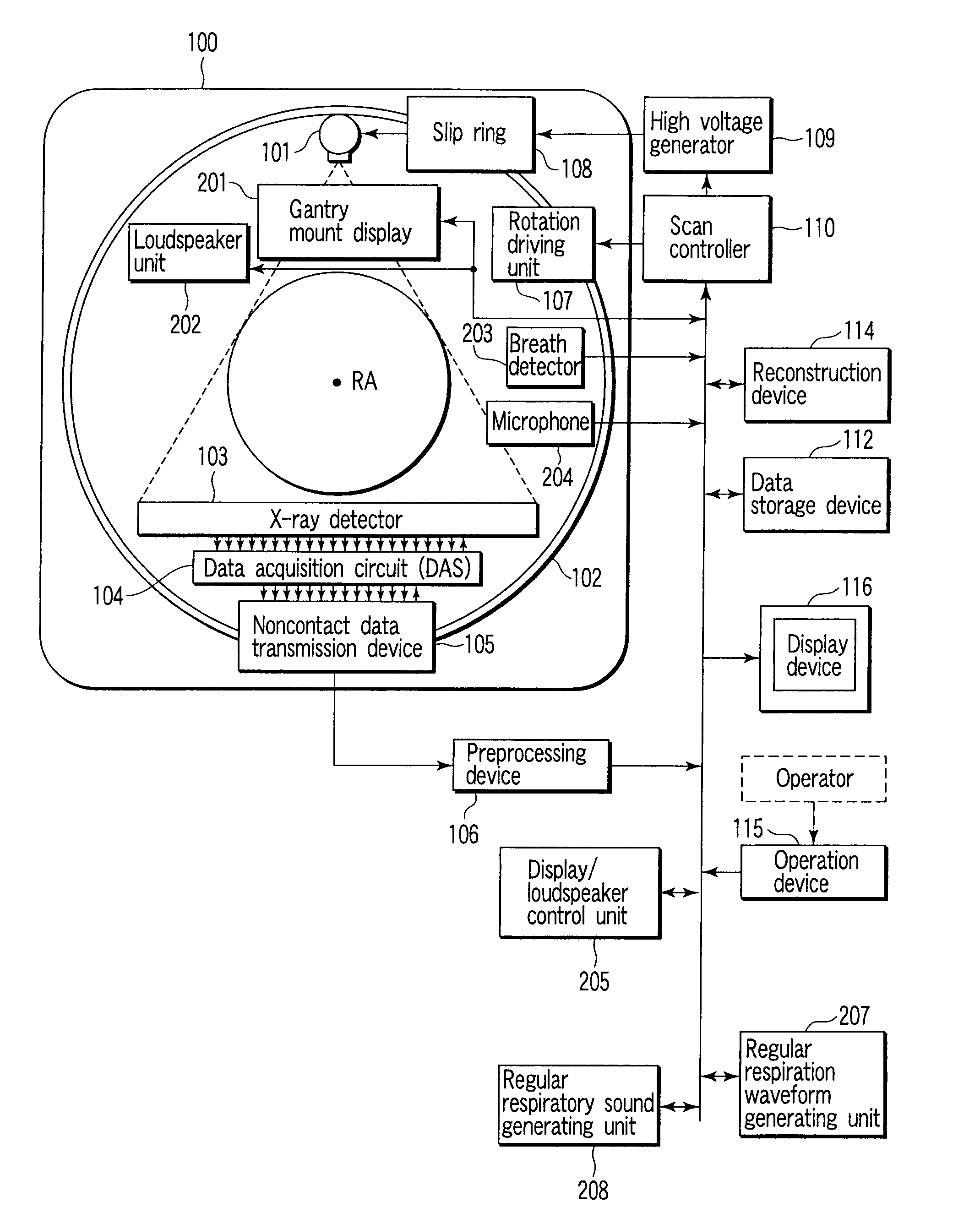
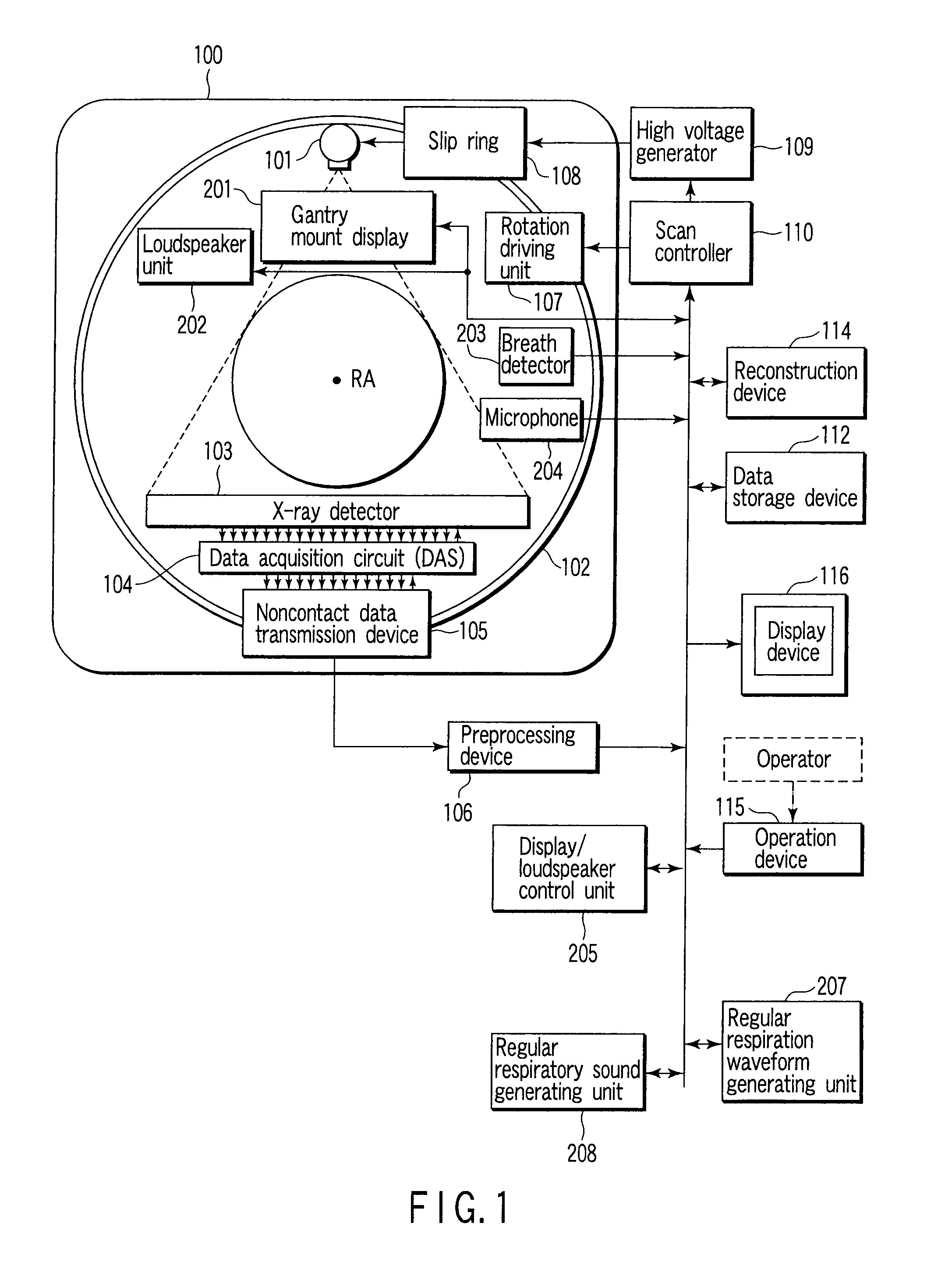
An X-ray computed tomographic apparatus includes a gantry 100 including an X-ray tube 101 which generates X-rays and an X-ray detector 103 which detects X-rays transmitted through a subject to be examined, a reconstruction device 114 which generates tomogram data on the basis of an output from the X-ray detector, a breath detector 203 which detects a respiration waveform representing a temporal change in respiration index value associated with the subject, a regular respiration waveform generating unit 207 which generates a respiration waveform with a regular respiration cycle which originates from the detected respiration waveform, and a gantry mount display 201 which displays the generated regular respiration waveform.

Description

CROSS-REFERENCE TO RELATED APPLICATIONS[0001]This application is a division of and claims the benefit of priority under 35 U.S.C. §120 from U.S. Ser. No. 11 / 870,633 filed Oct. 11, 2007, and claims the benefit of priority under 35 U.S.C. §119 from Japanese Patent Application Nos. 2006-277886 filed Oct. 11, 2006; No. 2006-282844 filed Oct. 17, 2006; and No. 2006-282845 filed Oct. 17, 2006, the entire contents of each of which are incorporated herein by reference.BACKGROUND OF THE INVENTION[0002]1. Field of the Invention[0003]The present invention relates to an X-ray computed tomographic apparatus, breath instruction apparatus, and medical imaging apparatus which generate medical images by imaging a subject to be examined.[0004]2. Description of the Related Art[0005]Stable breathing is indispensable for improving image quality by reducing artifacts. Conventionally, in order to stabilize breathing motion, an operator gives a subject to be examined spoken instructions from an operator ro...

Claims


Application Information

Patent Timeline
Patent Type & Authority Applications(United States)
IPC IPC(8): H05G1/60
CPCA61B5/08A61B5/721A61B6/541A61B6/032A61B6/5264A61B5/7285G01R33/283
Inventor NAKAMURA, HITOSHITSUYUKI, MASAHARUMAEDA, TATSUOXIAO, SHAN SHAN
Owner TOSHIBA MEDICAL SYST CORP
Learn More