Ultrasonic strain imaging device with selectable cost-function

a strain imaging and cost-function technology, applied in the field of ultrasonic imaging, can solve problems such as the possibility and the management of computational burden, and achieve the effect of improving images and inherent tension in matching kernels

Active Publication Date: 2010-04-29
WISCONSIN ALUMNI RES FOUND
View PDF27 Cites 27 Cited by
  • Summary
  • Abstract
  • Description
  • Claims
  • Application Information

AI Technical Summary

Benefits of technology

[0006]The present inventors have recognized that there is an inherent tension in matching kernels of the pre-deformation and post-deformation images between restoration of signal coherence (high correlation between matching kernels) and correctness of the matching with respect to other matches (continuity or limited tissue mobility), and that this tension may best be resolved using mathematical optimization where cost functions allow different trade-offs between correlation and continuity for different tissue types and imaging situations. By allowing this trade-off to be varied, for example, based on a priori knowledge about the imaging situation, improved images may be obtained.

Problems solved by technology

Normally the size of the search window must be sufficient to accommodate likely tissue displacements between the pre-deformation and post-deformation images, but must also be limited to manage the computational burden of matching points with each other and to reduce the chance of possible false matches that violate a priori assumptions about limited mobility of a tissue continuum reacting to external mechanical stimuli.

Method used


Image

  • Ultrasonic strain imaging device with selectable cost-function
  • Ultrasonic strain imaging device with selectable cost-function
  • Ultrasonic strain imaging device with selectable cost-function

Examples


Embodiment Construction

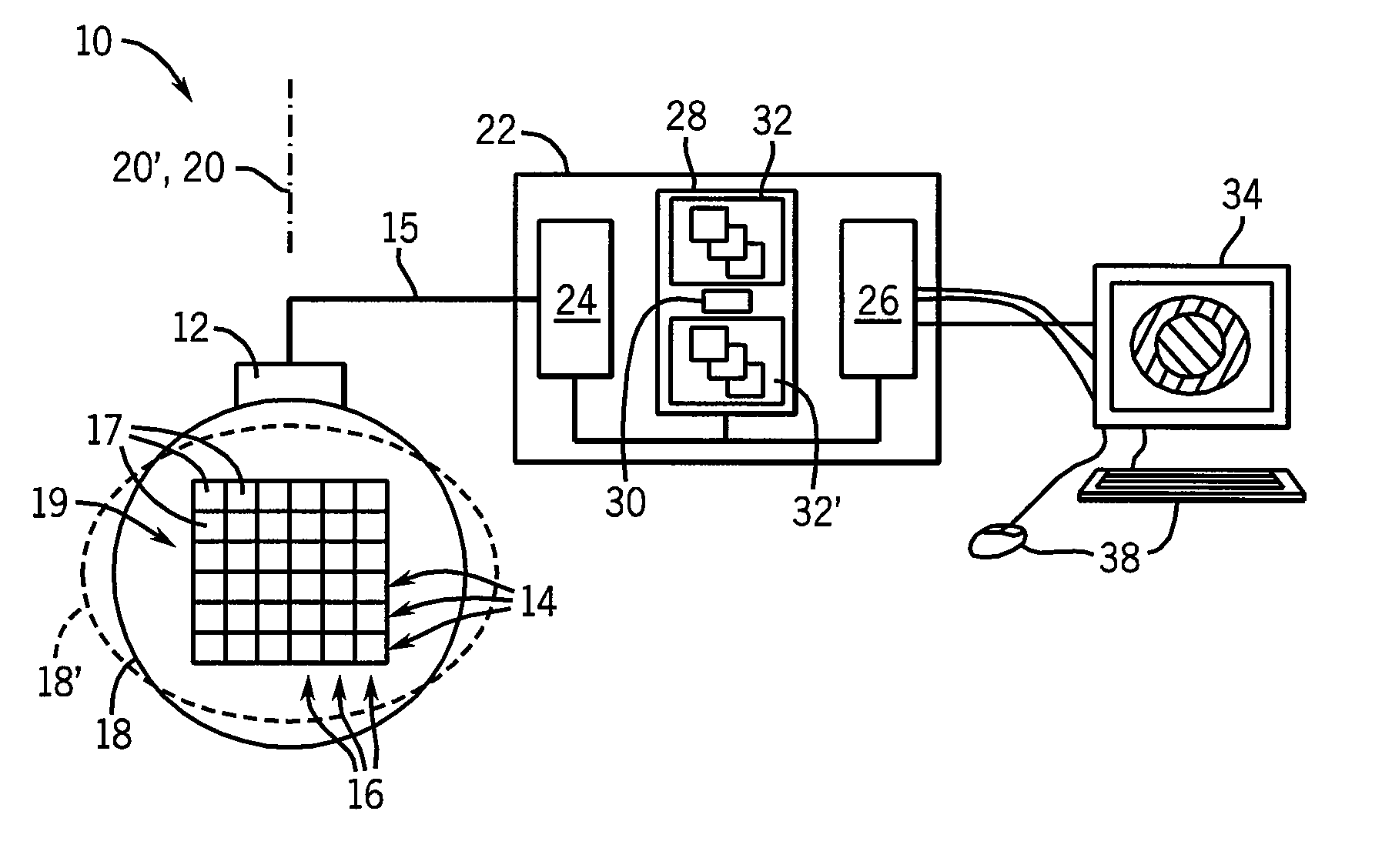
[0041]Referring now to FIG. 1, an elasticity imaging machine 10 of the present invention includes an ultrasonic array transducer 12 that may transmit and receive ultrasonic signals along a propagation axis 20 to acquire ultrasonic echo data 15 at corresponding volume elements 17 throughout a region of interest 19 in the tissue 18.

[0042]The echo data 15, and its corresponding volume elements 17, may both be identified by logical rows 14 and columns 16, wherein the rows 14 are generally echo data 15 or volume elements 17 extending perpendicularly to the propagation axis 20, and the columns 16 are generally echo data 15 or volume elements 17 extending parallel to the propagation axis 20. These terms should be understood generally to describe data acquired through a variety of ultrasonic acquisition geometries including those which provide for fan beams of ultrasound and the like, and therefore not be limited to strictly rectilinear rows and columns.

[0043]In addition to transmitting and...


PUM


Abstract

An elasticity measuring system determines tissue displacement between a pre-deformation and post-deformation image by a matching process using a cost function accepting as its arguments continuity of the tissue motion and correlation of the tissue in making the block matching. The invention allows the selection among different cost functions for different imaging situations or tissue types, to provide improved displacement calculations using a priori knowledge about the tissue and structure of tissue interfaces or information derived during the scanning process.

Description

STATEMENT REGARDING FEDERALLY SPONSORED RESEARCHCROSS-REFERENCE TO RELATED APPLICATIONSBACKGROUND OF THE INVENTION[0001]The present invention relates to ultrasonic imaging and, in particular, to an improved method and apparatus for calculating material displacement used to produce elasticity images including local strain, modulus and Poison' ratio images.[0002]Ultrasonic elasticity imaging produces an image showing the elastic parameters of the material being measured. When used in medicine, elasticity imaging is analogous to palpation by a physician, that is, the pressing of tissue by the physician to feel differences in elasticity of underlying structures.[0003]In a common form of elasticity imaging, two separate ultrasonic images are obtained, the first image with tissue in an undeformed state relative to the second image (“pre-deformation”) and the second image with the tissue in a deformed state (“post deformation”). The two images are analyzed to deduce the amount of displacem...

Claims


Application Information

Patent Timeline
Patent Type & Authority Applications(United States)
IPC IPC(8): A61B8/00
CPCA61B8/485A61B8/08
Inventor JIANG, JINGFENGHALL, TIMOTHY J.
Owner WISCONSIN ALUMNI RES FOUND
Why Eureka
Try Eureka