

Inhibition of autophagy genes in cancer chemotherapy
a cancer and autophagy technology, applied in the field of cancer chemotherapy, can solve the problems of morbidity worldwide and achieve the effect of reducing non-specificity and high selectivity
- Summary
- Abstract
- Description
- Claims
- Application Information
AI Technical Summary
Benefits of technology
Problems solved by technology
Method used
Image

Examples
example 1
[0105]A FACS based strategy coupled with quantitative mRNA and protein analysis is employed to quantify the effect of inhibition of autophagy at the molecular level in cancer treatment models. In addition, siRNA based knockdown is used to disrupt autophagy genes thereby allowing autophagy to be assessed selectively.
[0106]The results indicate that inhibition of autophagy in tamoxifen treated breast carcinoma cells leads to an increase in cell death.
[0107]Materials and Methods
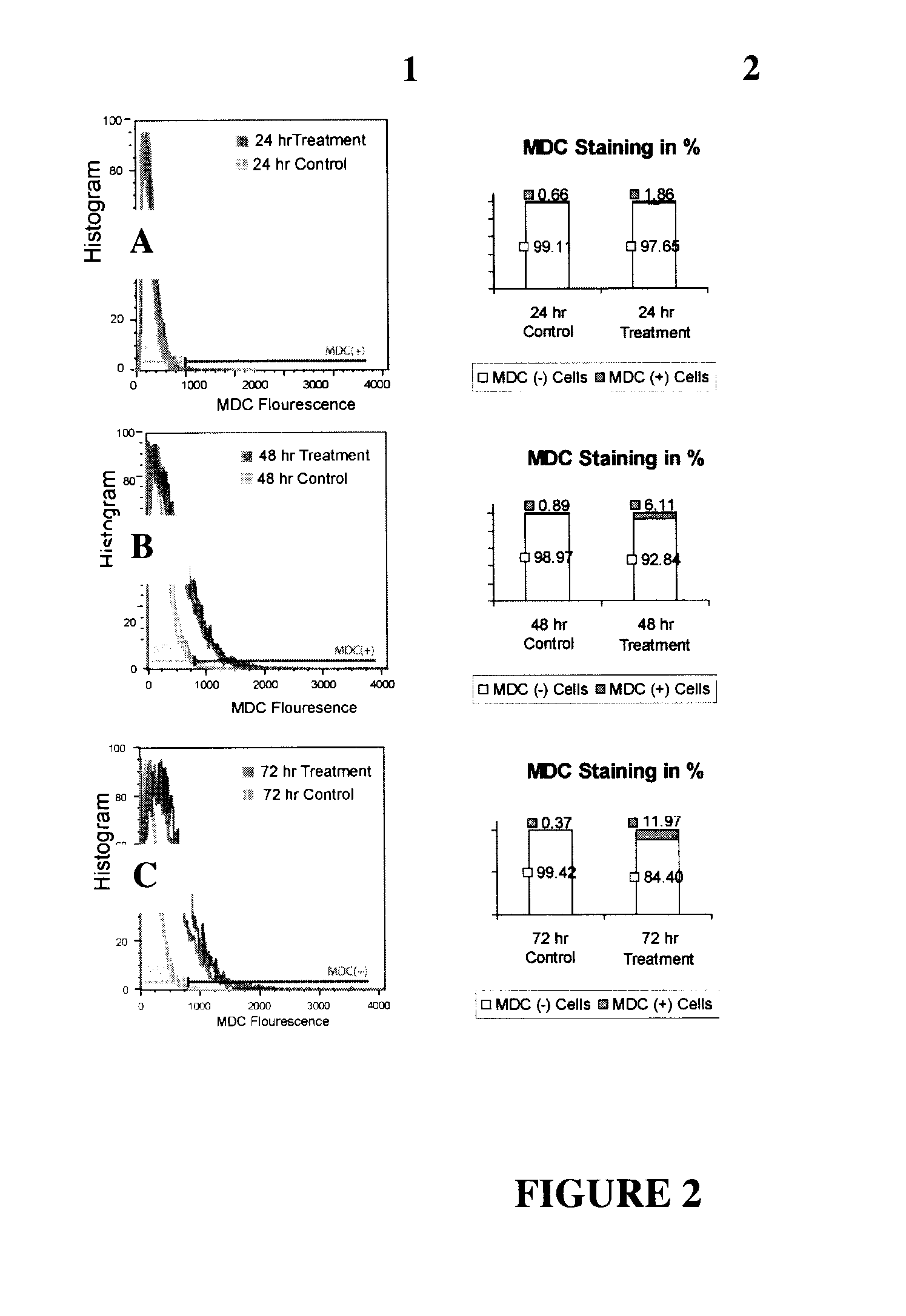
[0108]Atg gene-siRNA in breast carcinoma cell line: The human MCF7 breast carcinoma cell line is used in these studies for several reasons. First, MCF7 cells undergo autophagy following treatment with tamoxifen (9) and irradiation (11). Second, monodansylcadaverine (MDC) staining in tamoxifen-treated MCF7 cells correlates with autophagic activity (13) and thus provides a useful autofluorescent marker that can be used in addition with the autophagosome marker GFP-LC3 and the lysosomal stain LysoTracker® Green (LT-...
example 2
[0124]MDC (monodansylcadaverine): MDC is a lysosomotropic agent which preferentially labels autophago-lysosomes and some autophagosomes. It is believed that the specificity of MDC staining of autophagolysosomes depends on some kind of interaction between MDC and the lipid molecules, which are abundant in autophagolysosomes.
[0125]MAP1LC3B & Beclin 1: MAP1LC3B is the human orthologue of Yeast autophagy gene ATG 8. ATG 8 is essential for the elongation of the “isolation membrane” which makes up the structure of the autophagosome and serves as an indicator for autophagy. Our analysis reveals that the expression level of MAP1LC3B correlates with an increase in autophagy as detected by an increase in MDC signal. Beclin 1 is the human orthologue of Yeast Autophagy gene ATG 6 and has been shown to be a regulator of mammalian autophagy.
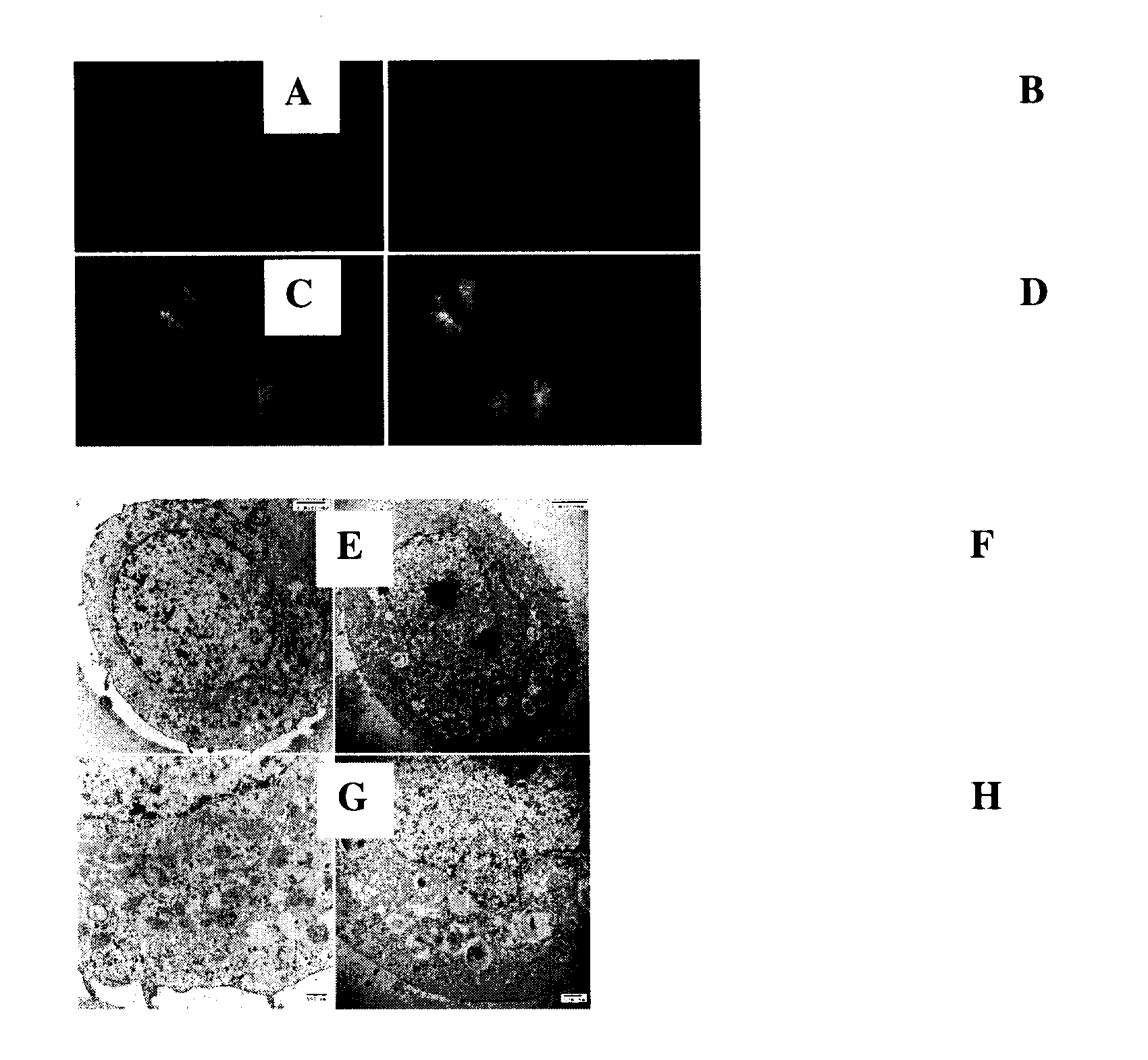
[0126]FIG. 8. Tamoxifen induces autophagy in MCF-7 cells: A. MCF-7 control cells. B. MCF-7 cells treated with 10-6 M tamoxifen. In panels A, B, & D, cells are...
example 3
[0133]FIG. 12. Measuring MDC-positive structures using the INCell Analyzer. Panel A is untreated (control) MCF7 cells, Panel B is 5 uM tamoxifen treated MCF7 cells, and Panel C shows the InCell Analyzer program running the “cell inclusion” algorithm to identify the number of MDC-positive inclusions per cell. Panel D illustrates the results of such an analysis conducted on triplicate samples.
[0134]FIG. 13. Autophagy detection by GFP-LC3 localization. Left: Diffuse GFP staining correlates with no autophagy (GFP-LC3 is cytoplasmic). Right: Punctate GFP staining correlates with autophagy (GFP-LC3 is bound to autophagosomal membranes; arrow).
[0135]FIG. 14. Effect of siRNA inhibition on Tamoxifen treated MCF-7 cells by WST-1 Assay. Panels A1, A2, and A3 are beclin 1, hAtg5 and hATG7 siRNA treated cells on Day 05 of Tamoxifen treatment. X-axis denotes untreated, 1×10⁻⁶ M and 5×10⁻⁶ M tamoxifen treated MCF-7 cells. Each siRNA panel contains the following: No siRNA (no treatment), negative c...
PUM
| Property | Measurement | Unit |
|---|---|---|
| Composition | aaaaa | aaaaa |
| Chemotherapeutic properties | aaaaa | aaaaa |
Abstract
Description
Claims
Application Information