Genomic classification of malignant melanoma based on patterns of gene copy number alterations
a gene copy number and malignant melanoma technology, applied in the field of gene copy number alterations for malignant melanoma, can solve the problems of not representing the genetic heterogeneity of the parent tumor, difficult to predict clinical outcome of individual melanoma, poor response in clinical trials to agents,
- Summary
- Abstract
- Description
- Claims
- Application Information
AI Technical Summary
Benefits of technology
Problems solved by technology
Method used
Image
Examples
example 1
CGH Data of Cell Lines and Tumor Tissue Samples
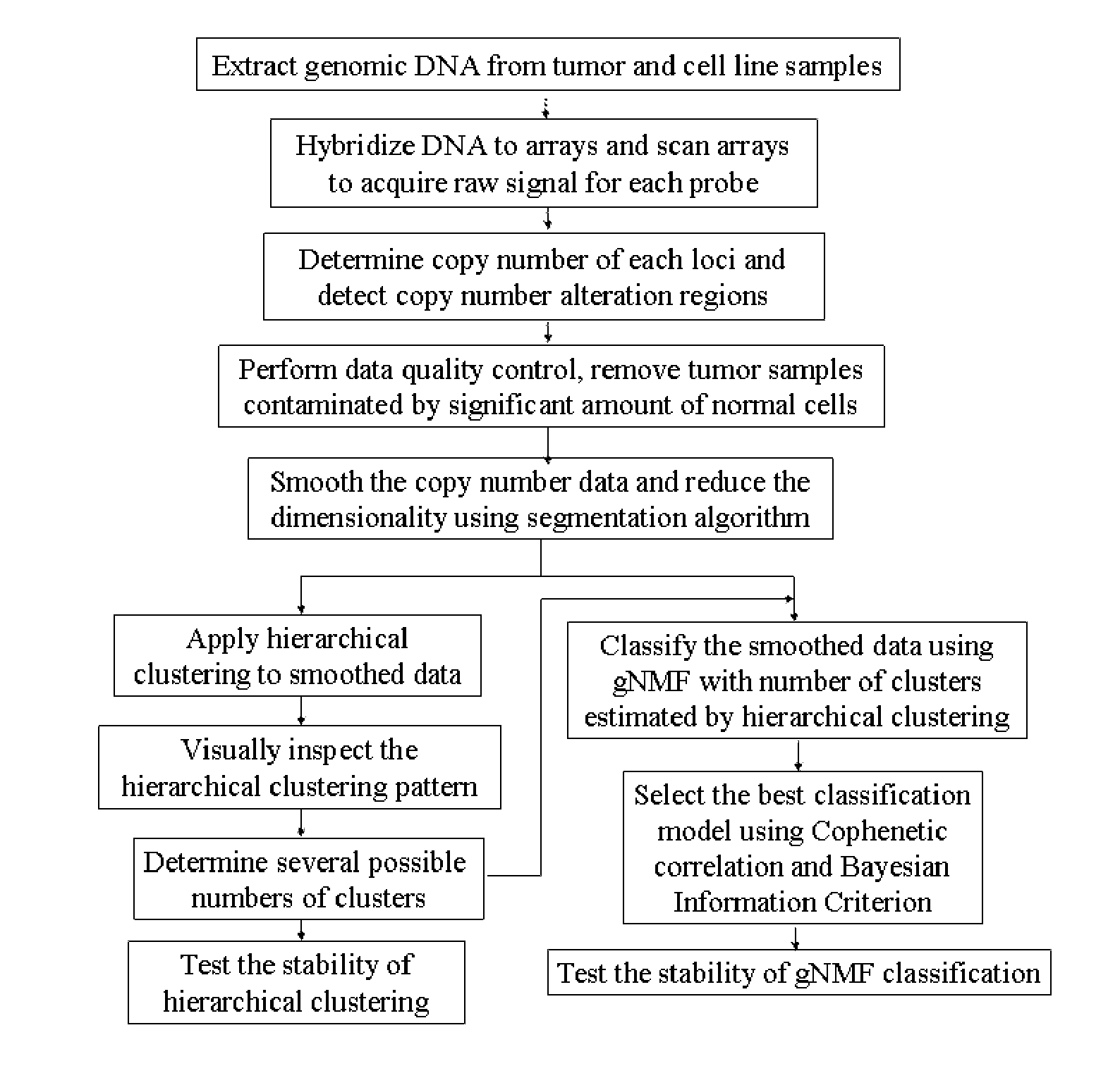
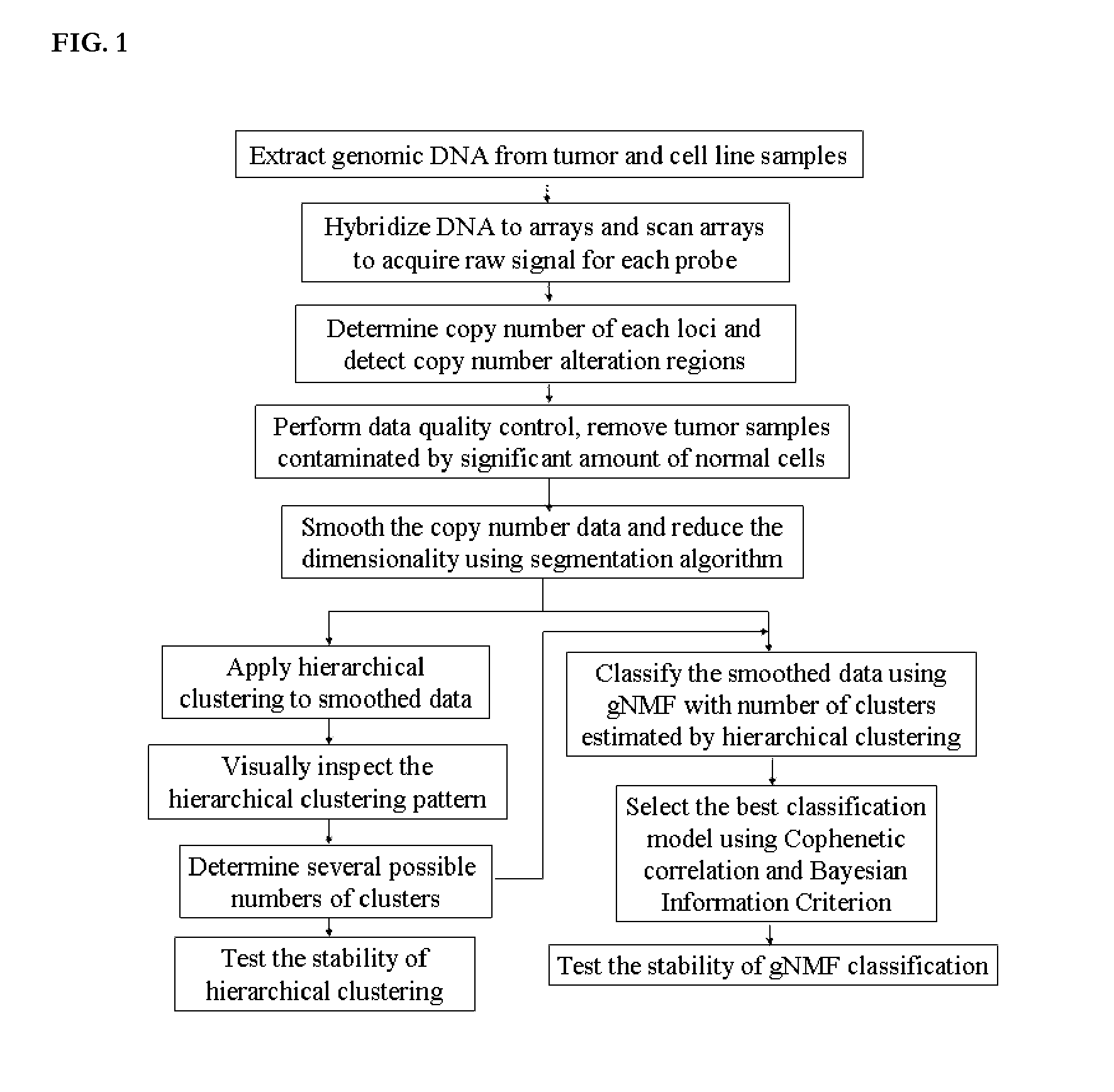
[0219]The inventors gathered CGH data for 30 melanoma cell lines and 109 melanoma short-term cultures from various published sources (Greshock et al., 2007; Lin et al., 2008) to establish the melanoma classification model. The sources of cell lines used in this study are listed in Table 1. These data had been acquired using Affymetrix's GENECHIP® Mapping 250K STY SNP arrays, following the manufacturer's instructions.
[0220]Copy number data can also be acquired using other SNPs or CGH microarray platforms, such as other versions of AFFYMETRIX® SNPs microarrays, Agilent aCGH microarrays (Agilent, Inc., Santa Clara, Calif.), ILLUMINA® microarrays (Illumina, Inc., San Diego, Calif.), and NIMBLEGEN® aCGH microarrays (Nimblegen, Inc., Madison, Wis.).
example 2
Step 2: Copy Number Determination and Detection of Copy Number Alterations
[0221]Genomic Suite software (version 6.08.0103) (Partek; St. Louis, Mo.) was used for low-level processing of the data to determine the copy numbers of each locus and define regions of copy number alteration. CEL files containing signals for all SNPs probes were loaded into the software, and copy numbers were calculated by comparing the signal intensities for tumor or cell line samples to those for a reference set of 90 normal female tissue samples, corrected to a baseline of 2. The reference set can also consist of other sets of normal samples, or paired normal tissues from the same patients of the tumor samples, measured by the same microarray platform.
[0222]The resulting probe-level copy number data were segmented, and the copy number alteration regions were detected in each sample. Specifically, probe-level copy numbers were segmented into regions using the following control parameters: (i) a region must ...
example 3
Step 3: Data Quality Control
[0224]Tumor samples may contain a significant percentage of normal cells that dilute the signal of copy number alteration present in the tumor cells. A machine learning algorithm to capture the difference between copy number patterns of tumor and normal samples was developed and then used to identify and eliminate normal contaminated samples from further analyses. First, a subset of samples with the highest number of copy number alteration regions and a set of normal samples were selected. These two groups of samples were used to train a machine learning algorithm (Random Forest: RF (Breiman, 2001)) to classify normal and tumor samples by tuning the parameters to best represent the difference between tumor and normal samples. Second, the trained classifier algorithm was applied to the rest of samples; the classifier assigned a score to each sample, where the score represented the probability of the sample being contaminated by normal cells. Samples that h...
PUM
| Property | Measurement | Unit |
|---|---|---|
| Fraction | aaaaa | aaaaa |
| Fraction | aaaaa | aaaaa |
| Biological properties | aaaaa | aaaaa |
Abstract
Description
Claims
Application Information
 - R&D
- Intellectual Property
- Life Sciences
- Materials
- Tech Scout
- Unparalleled Data Quality
- Higher Quality Content
- 60% Fewer Hallucinations
Browse by: Latest US Patents, China's latest patents, Technical Efficacy Thesaurus, Application Domain, Technology Topic, Popular Technical Reports.
© 2025 PatSnap. All rights reserved.Legal|Privacy policy|Modern Slavery Act Transparency Statement|Sitemap|About US| Contact US: help@patsnap.com


