
Removable Vena Cava Filter

a filter and vena cava technology, applied in the field of medical devices, can solve the problems of thrombosis posing a serious risk of pulmonary embolism, filters generally have not been considered to be removable from patients, and downstream embolism or embolization, so as to avoid interference with blood flow, reduce cross-sectional area, and reduce the diameter

Active Publication Date: 2010-06-24
COOK INC
View PDF99 Cites 22 Cited by
  • Summary
  • Abstract
  • Description
  • Claims
  • Application Information

AI Technical Summary

Benefits of technology

[0010]The present invention provides a vena cava filter comprising struts configured to align the filter about the center axis of a blood vessel and minimize engagement with the blood vessel. The filter comprises a plurality of primary struts, each of which having a first end. A hub axially connects the first ends of the struts to define a central axis of the filter. Each primary strut has a curved member extending from the central axis. Each curved member terminates at an anchoring hook to engage the blood vessel at a first axial plane and secure the filter in the blood vessel. Each anchoring hook includes a barb formed at an angle relative to the strut to allow a removal sheath to be advanced over the filter and allow the hooks to be removed straight away from the vessel wall, resulting in minimal vessel damage. The filter further comprises a plurality of secondary struts. Each secondary strut is connected to one of the curved members and extends therefrom to a free end for engaging the blood vessel at a second axial plane, aligning the filter in the blood vessel.
[0011]In one embodiment, a set of at least two secondary struts are connected to the curved member of one primary strut. The set of secondary struts extend radially from each side of the primary strut, forming a netting configuration of the filter. In another embodiment, one secondary strut is connected to the curved member of one primary strut. The secondary strut extends from the primary strut and is in radial alignment with the primary strut, avoiding interference with blood flow.
[0012]In a collapsed configuration, the vena cava filter occupies a reduced diameter, since the hub is the origin to only primary struts. In an expanded configuration, the hub occupies a reduced cross-sectional area. As a result, interference with blood flow is lessened in the vena cava.
[0013]In an expanded configuration, the vena cava filter occupies a reduced length, since the secondary struts merely extend within the axial length of the primary struts. As a result, the filter can more easily be placed in the vena cava of a patient, lessening the risk of interference in the femoral and renal veins.

Problems solved by technology

During such medical conditions, the need for filtering devices arises due to the likelihood of thrombosis in the peripheral vasculature of patients wherein thrombi break away from the vessel wall, risking downstream embolism or embolization.
For example, depending on the size, such thrombi pose a serious risk of pulmonary embolism wherein blood clots migrate from the peripheral vasculature through the heart and into the lungs.
For example, filters generally have not been considered removable from a patient due to the likelihood of endotheliosis of the filter during treatment.
Moreover, conventional filters commonly become off-centered or tilted with respect to the hub of the filter and the longitudinal axis of the vessel in which it has been inserted.
As a result, the filter including the hub and the retrieval hook engage the vessel wall along their lengths and potentially become endothelialized therein.
In the event of this occurrence, there is a greater likelihood of endotheliosis of the filter to the blood vessel along a substantial length of the filter wire.
As a result of the two levels, removal of the filter from the blood vessel becomes impossible or at least difficult.
If the lower part of the filter extends into the femoral veins, filtering effectiveness will be compromised.
Moreover, it is not desirable to have filter wires crossing the origin of the renal veins, since the filter wires may interfere with the flow of blood from the kidneys.
The resulting diameter of this bundle of filter wires results in a filter that may be too large for easy placement and becomes an obstacle to blood flow in the vena cava.

Method used


Image

  • Removable Vena Cava Filter
  • Removable Vena Cava Filter
  • Removable Vena Cava Filter

Examples


Embodiment Construction

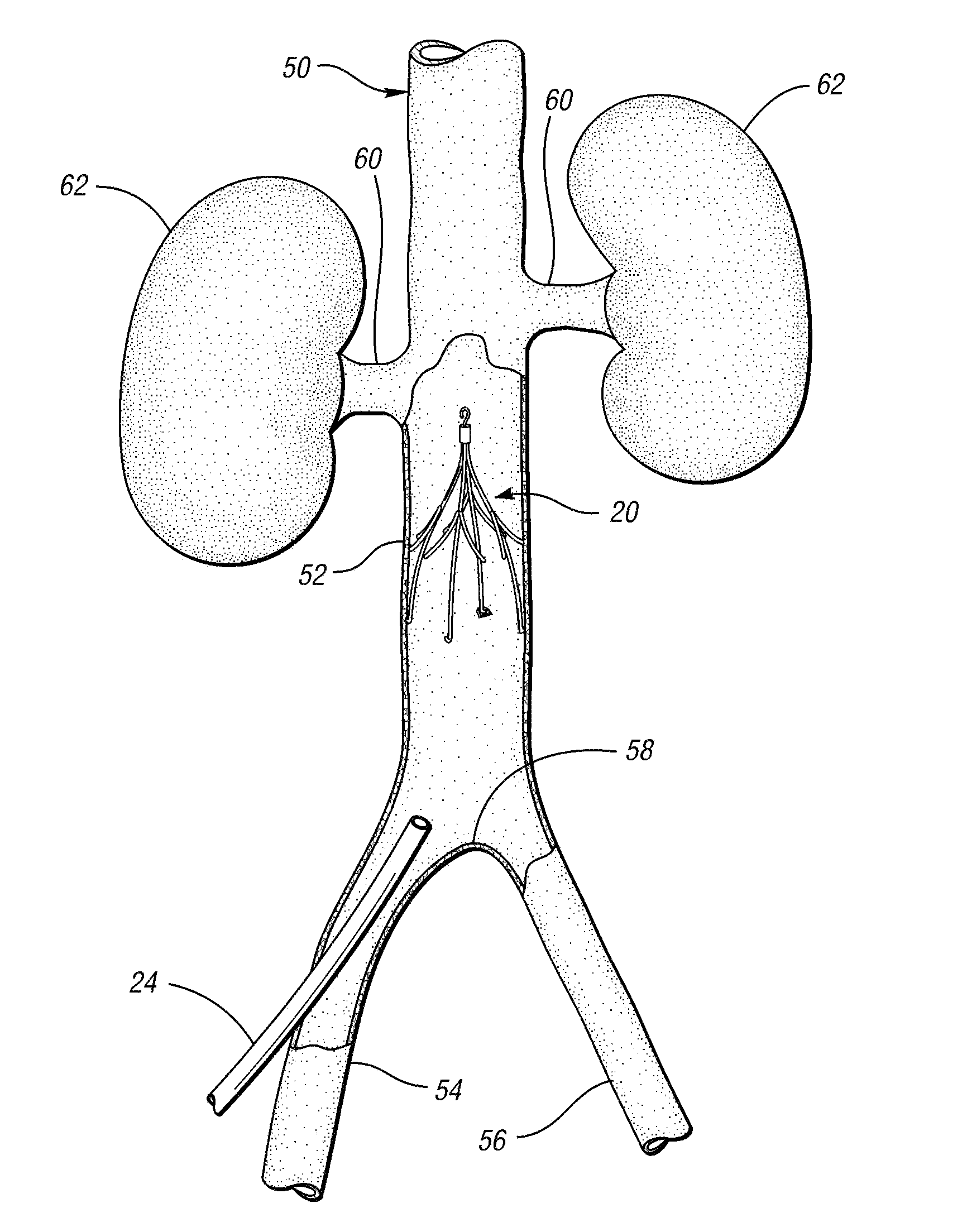
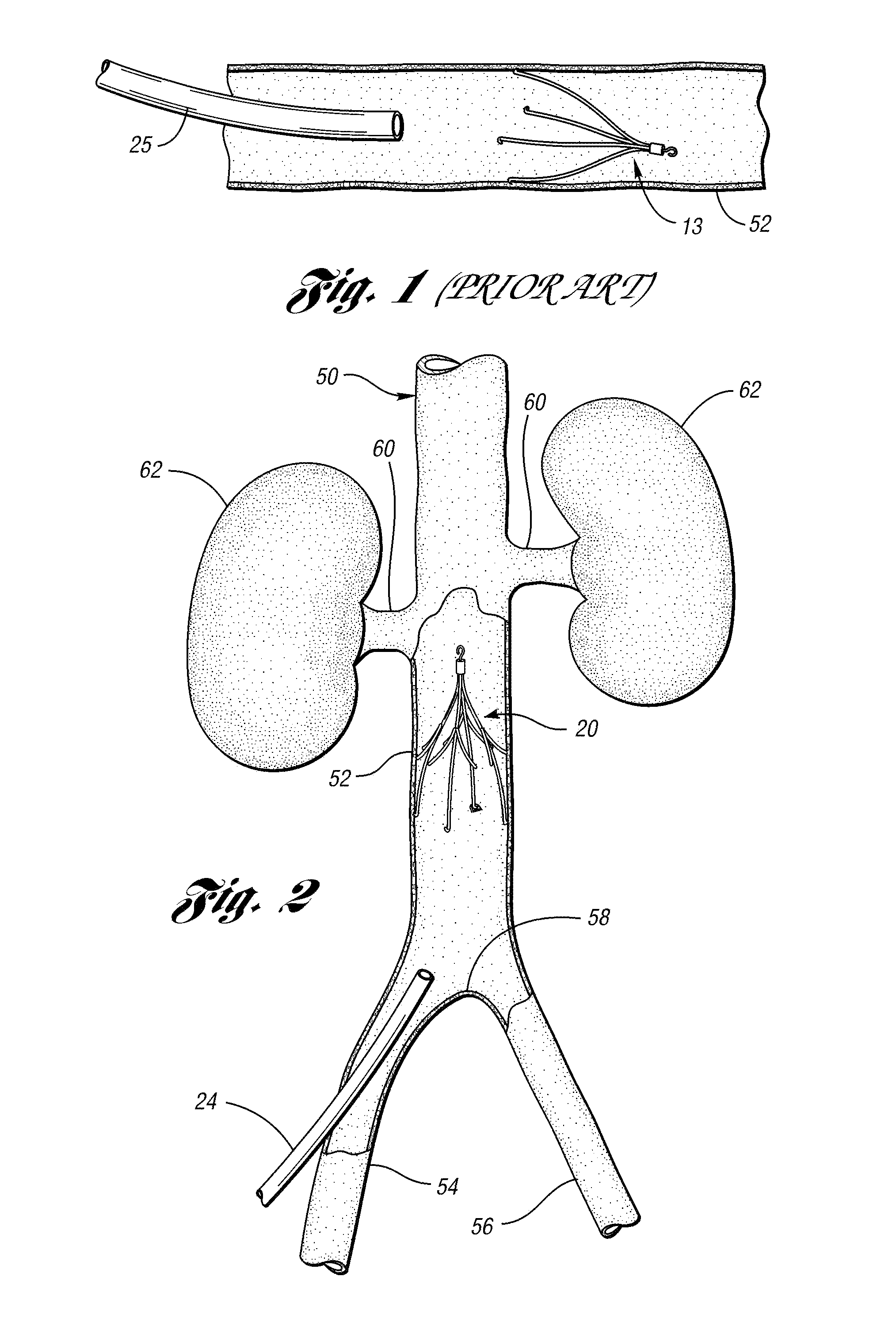
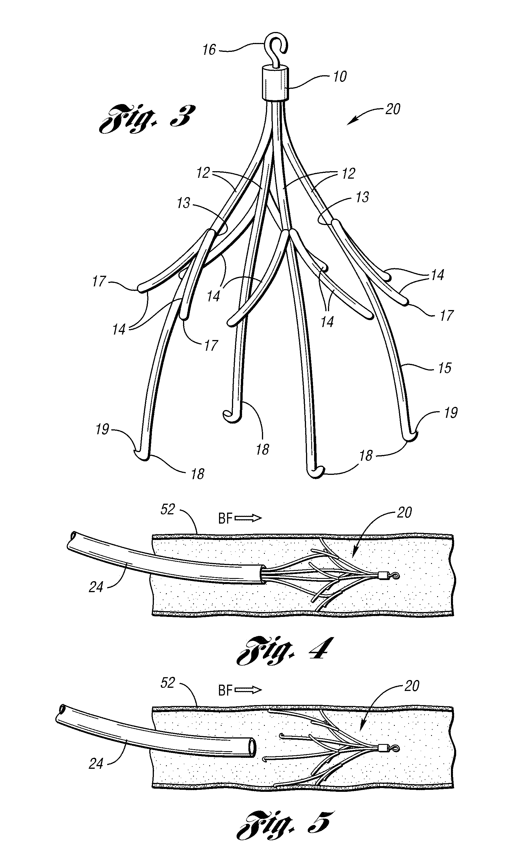
[0025]In accordance with a first embodiment of the present invention, FIG. 2 illustrates a vena cava filter 20 implanted in the vena cava 50 for the purpose of lysing or capturing thrombi carried by the blood flowing through the femoral veins 54, 56 toward the heart and into the pulmonary arteries. As shown, the femoral veins from the legs merge at juncture 58 into the vena cava 50. The renal veins 60 from the kidneys 62 join the vena cava 50 downstream of juncture 58. The portion of the vena cava 50, between the juncture 58 and the renal veins 60, defines the inferior vena cava 52 in which the vena cava filter 20 has been percutaneously deployed through one of the femoral veins 54. Preferably, the vena cava filter 20 has a length smaller than the length of the inferior vena cava 52. If the lower part of the filter extends into the femoral veins, filtering effectiveness will be compromised and if the filter wires cross over the origin of the renal veins the filter wires might interf...


PUM


Abstract

A removable filter for capturing thrombi in a blood vessel. The filter comprises a plurality of primary struts having first ends connected to each other to define a central axis of the filter. Each primary strut has a curved member extending from the central axis and terminates at an anchoring hook to engage the blood vessel at a first axial plane. The filter further comprises a plurality of secondary struts connected to the curved members of the primary struts and extending therefrom to a free end at a second axial plane to centralize the filter in the blood vessel.

Description

CROSS-REFERENCE TO RELATED APPLICATIONS[0001]This application is a divisional of U.S. patent application Ser. No. 10 / 776,721, filed Feb. 11, 2004, entitled “REMOVABLE VENA CAVA FILTER”, which claims the benefit of U.S. Provisional Patent Application Ser. No. 60 / 446,711, filed Feb. 11, 2003, entitled, “REMOVABLE VENA CAVA CLOT FILTER,” each of which is hereby incorporated by reference in its entirety.BACKGROUND OF THE INVENTION[0002]The present invention relates to medical devices. More particularly, the invention relates to a removable vena cava clot filter that can be percutaneously placed in and removed from the vena cava of a patient.[0003]Filtering devices that are percutaneously placed in the vena cava have been available for over thirty years. A need for filtering devices arises in trauma patients, orthopedic surgery patients, neurosurgery patients, or in patients having medical conditions requiring bed rest or non-movement. During such medical conditions, the need for filteri...

Claims


Application Information

Patent Timeline
Patent Type & Authority Applications(United States)
IPC IPC(8): A61F2/01
CPCA61F2/01A61F2002/011A61F2230/008A61F2230/005A61F2230/0078A61F2002/016A61F2/0103A61F2/0105A61F2/011
Inventor OSBORNE, THOMAS A.
Owner COOK INC
Features
  • Generate Ideas
  • Intellectual Property
  • Life Sciences
  • Materials
  • Tech Scout
  • Unparalleled Data Quality
  • Higher Quality Content
  • 60% Fewer Hallucinations
Learn More