Seroconversion assays for detecting xenotropic murine leukemia virus-related virus

a technology of xenotropic murine leukemia and serum antibodies, which is applied in the field of serum antibody detection, can solve the problem that the finding has not been extended to the detection of seroconversion antibodies against xmrv in humans

Inactive Publication Date: 2010-07-01
WHITTEMORE PETERSON INST FOR NEURO IMMUNE DISEASE
View PDF2 Cites 10 Cited by
  • Summary
  • Abstract
  • Description
  • Claims
  • Application Information

AI Technical Summary

Benefits of technology

[0031]In additional embodiments, methods of the present teachings can further comprise selecting or modifying a treatment on the basis of the detection of antibody against XMRV in a sample from a subject. In various aspects, if antibody against XMRV is detected in the sample, the treatment can comprise administrating to the subject a therapeutically effective amount of an anti-viral compound. In some configurations, the antiviral compound can be, without limitation, a compound such as acyclovir, penciclovir (famciclovir), gancyclovir (g

Problems solved by technology

However, these findings have not been extended to detect

Method used


Image

  • Seroconversion assays for detecting xenotropic murine leukemia virus-related virus
  • Seroconversion assays for detecting xenotropic murine leukemia virus-related virus
  • Seroconversion assays for detecting xenotropic murine leukemia virus-related virus

Examples


example 1

[0040]This example illustrates direct detection of antibody against XMRV in plasma from CFS patients.

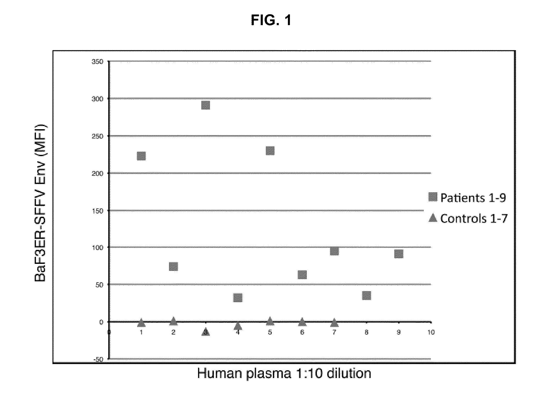
[0041]Our prior demonstration that infectious virus is present in both T and B lymphocytes from CFS patients is consistent with the tropism of other well-documented targets of human retroviral infection (B. J. Poiesz et al., Proc Natl Acad Sci USA 77, 7415 1980; J. C. Chemann et al., Antibiot Chemother 32, 48, 1983). We investigated whether XMRV stimulated an immune response in these patients, and developed an assay for detecting antibodies to XMRV ENV. This assay uses a murine pro B cell line, BAF-3 (control) and BAF-3 stably expressing SFFVgp55 ENV. In these experiments, Plasma from CFS patients or normal healthy controls was diluted 1:10, reacted with BaF3-ER or BaF3ERSFFV Env cells and analyzed by intracellular flow cytometry (IFC). FIG. 1 shows the difference in mean fluorescence intensity (MFI) between CFS and control plasma direct binding to BaF3ER-SFFV Env cells versus BaF3ER...

example 2

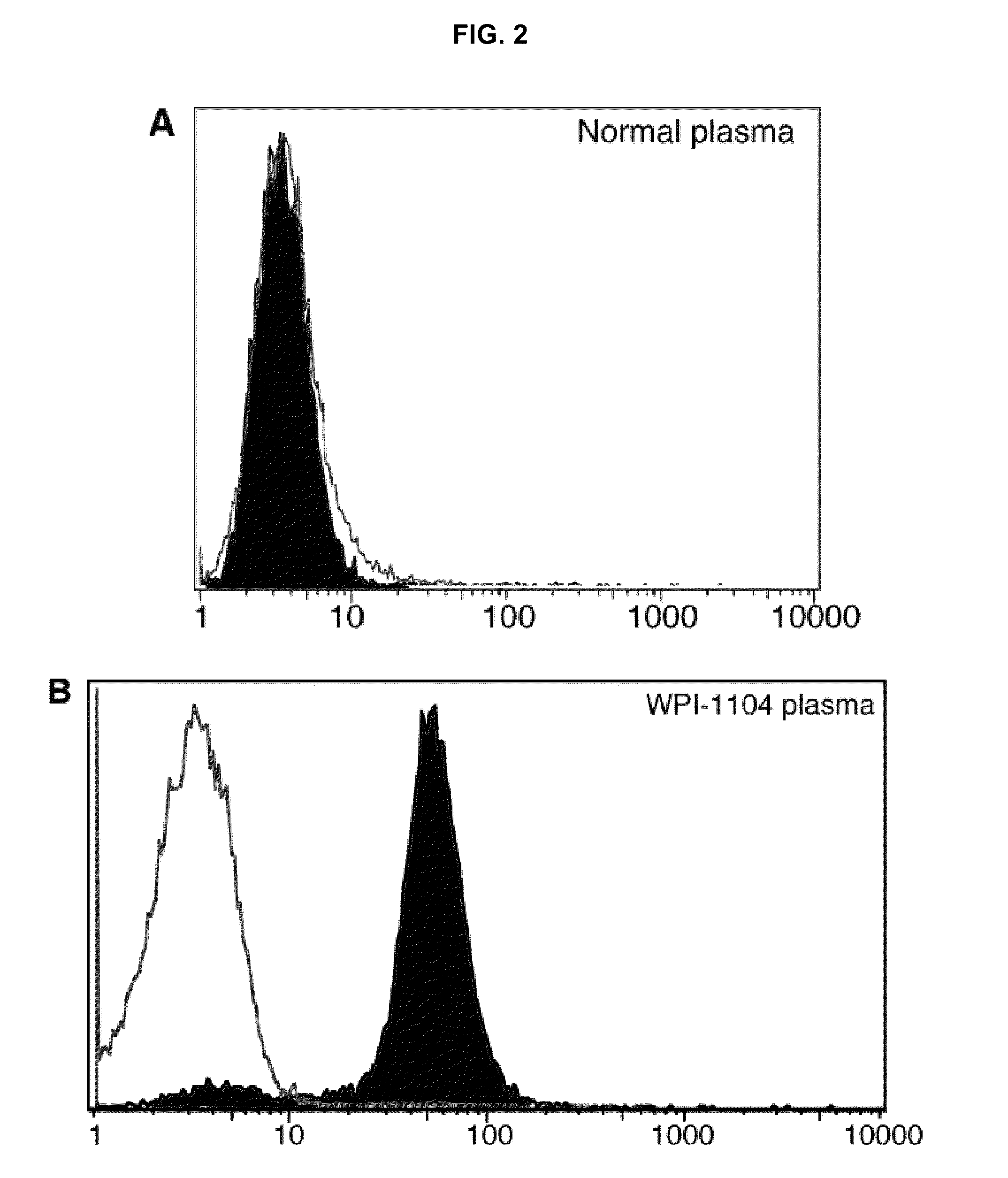
[0042]This example illustrates detection of antibody against XMRV in plasma from CFS patients. In these experiments, as illustrated in FIG. 2, samples of human plasma were assayed by flow cytometry for presence of antibody against XMRV using direct and competitive assays.

[0043]Direct assay of binding of normal plasma with BaF-3-SFFV ENV was negative (FIG. 2A). Furthermore, as shown in FIG. 2B, direct assay of binding of normal plasma with BaF-3 ER FC was also negative. However, a plasma sample from a patient who was previously diagnosed clinically with CFS (designated patient 1104 by the Whittemore Peterson Institute), was found to comprise antibody against XMRV: as shown in FIG. 2B, black area, a 1:10 dilution of plasma was positive in this assay. FIG. 2C illustrates direct binding of 2 μL 7C10 monoclonal anti SFFV Env antibody to BAF3-SFFV gp55 cell line (black area) vs. binding to BaF-3 ER control (light area). FIG. 2D illustrates competition for binding of 7C10 to BAF3-SFFV gp55...

example 3

[0044]This example illustrates detection of antibodies against XMRV in sera of patients, using direct and competitive assays (FIG. 3).

[0045]We investigated whether XMRV stimulates an immune response in CFS patients. For this purpose, we developed a flow cytometry assay that allowed us to detect antibodies to XMRV Env by exploiting its close homology to SFFV Env (Wolff, L., et al., Proc. Nat'l. Acad. Sci. USA 80: 4718-4722, 1983).

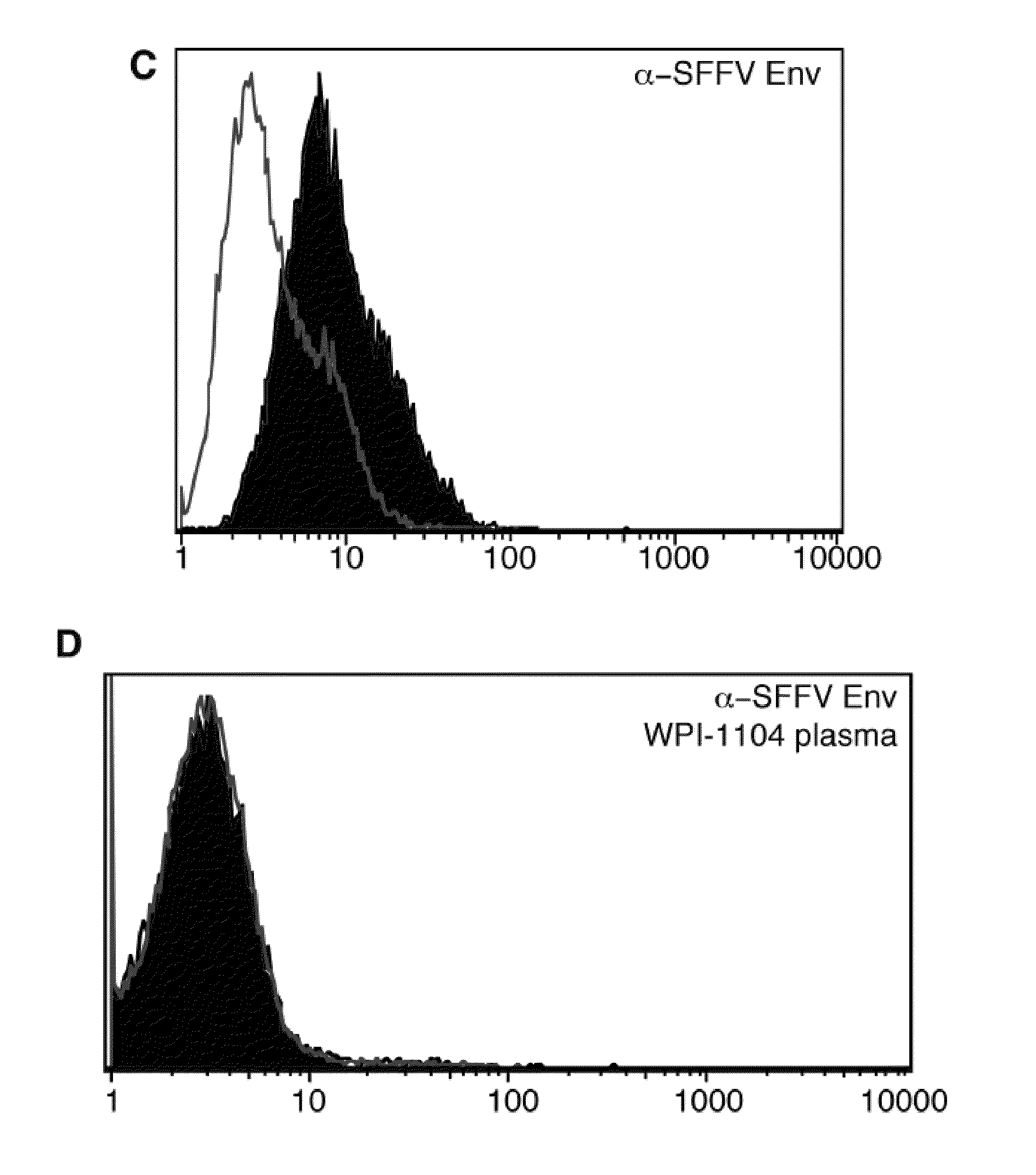
[0046]FIG. 3A illustrates no direct binding on BAF3ER control cells. Left panel: binding of human plasma at 1:10 dilution as detected by anti human IgG; right panel: no binding of anti-SFFV env monoclonal (7C10) at 1:10 dilution as detected using an anti rat IgG. Y3 rat hybridoma supernatant served as control. FIG. 3B illustrates direct binding on BAF3ER-SFFV Env cells. Left panel illustrates direct binding of human CFS from patient 1104 but not normal plasma at 1:10 dilution as detected by anti human IgG; right panel illustrates direct binding of anti-SFFV ...


PUM


Abstract

Methods of detecting, diagnosing, monitoring or managing an XMRV-related disease such as an XMRV-related neuroimmune disease such as chronic fatigue syndrome or an XMRV-related lymphoma such as mantle cell lymphoma in a subject are disclosed. These methods comprise determining presence, absence or quantity of antibodies against XMRV in a sample from a subject.

Description

CROSS-REFERENCE TO RELATED APPLICATIONS[0001]This application claims priority from U.S. Provisional Application Ser. No. 61 / 225,877 filed on Jul. 15, 2009, which is incorporated herein by reference in its entirety.GOVERNMENT INTERESTS[0002]This work was supported at least in part with funds from the federal government under U.S.P.H.S. Grant HHSN26120080001E and Grant NCI / NIH CA104943 awarded by the National Institutes of Health, and Grant W81XWH-07-1338 awarded by U.S. Department of Defense Prostate Cancer Research Program. The U.S. Government may have certain rights in the invention.INCORPORATION-BY-REFERENCE OF MATERIAL SUBMITTED IN COMPUTER READABLE FORM[0003]The Sequence Listing, which is a part of the present disclosure, includes a computer readable form and a written sequence listing comprising nucleotide and / or amino acid sequences of the present disclosure. The sequence listing information recorded in computer readable form is identical to the written sequence listing. The s...

Claims


Application Information

Patent Timeline
IPC IPC(8): C12Q1/70
CPCG01N33/56983G01N2333/15G01N2469/20
Inventor MIKOVITS, JUDY A.RUSCETTI, FRANCIS W.RUSCETTI, SANDRA K.
Owner WHITTEMORE PETERSON INST FOR NEURO IMMUNE DISEASE
Why Eureka
Try Eureka