

Tetrameric cytokines with improved biological activity
a technology of tetrameric cytokines and biological activity, which is applied in the direction of immunological disorders, drug compositions, peptides, etc., can solve the problems that the prognosis of ifn as a cancer treatment has been primarily hindered, and achieves the effects of improving efficacy, long infusion time, and low toxicity
- Summary
- Abstract
- Description
- Claims
- Application Information
AI Technical Summary
Benefits of technology
Problems solved by technology
Method used
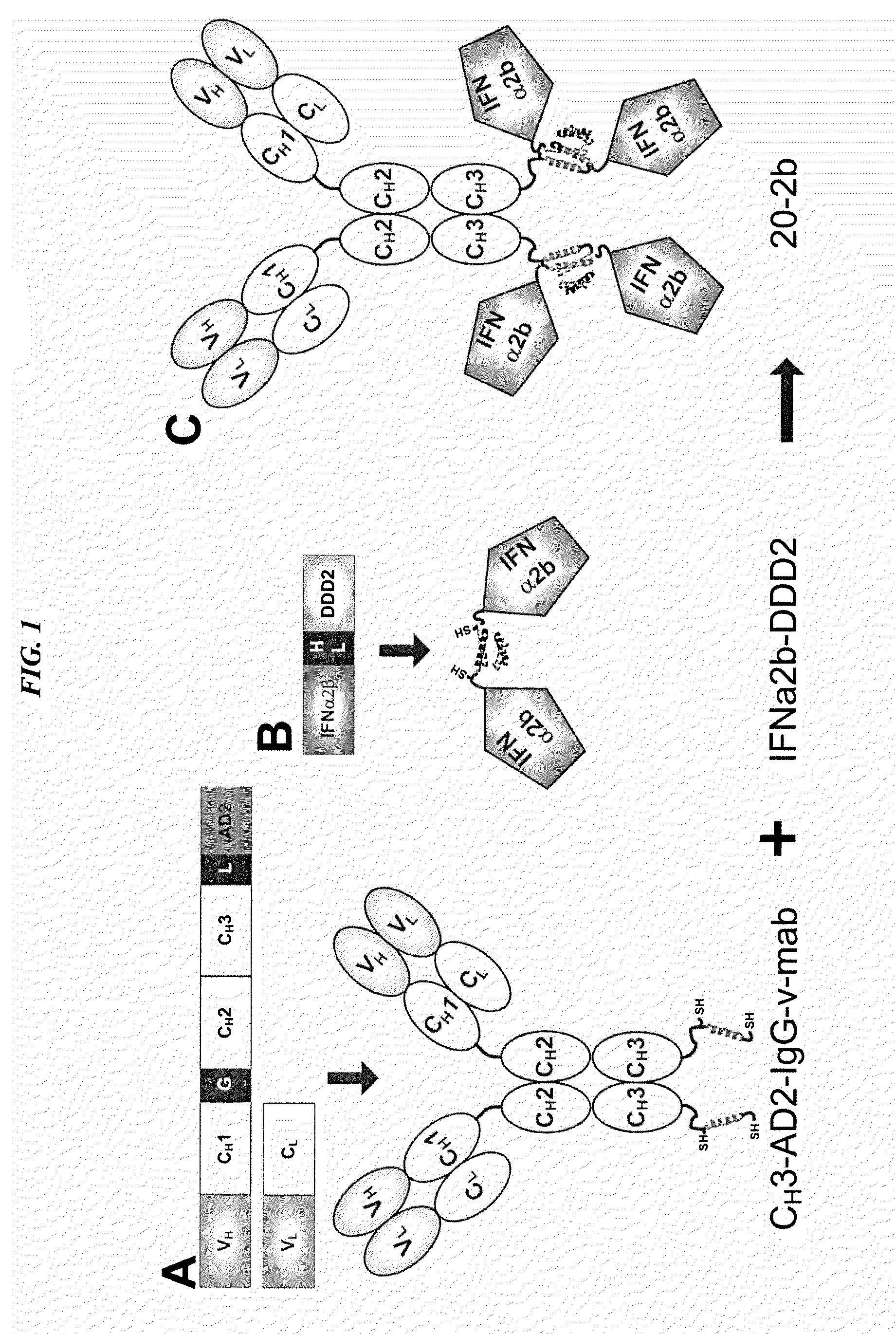
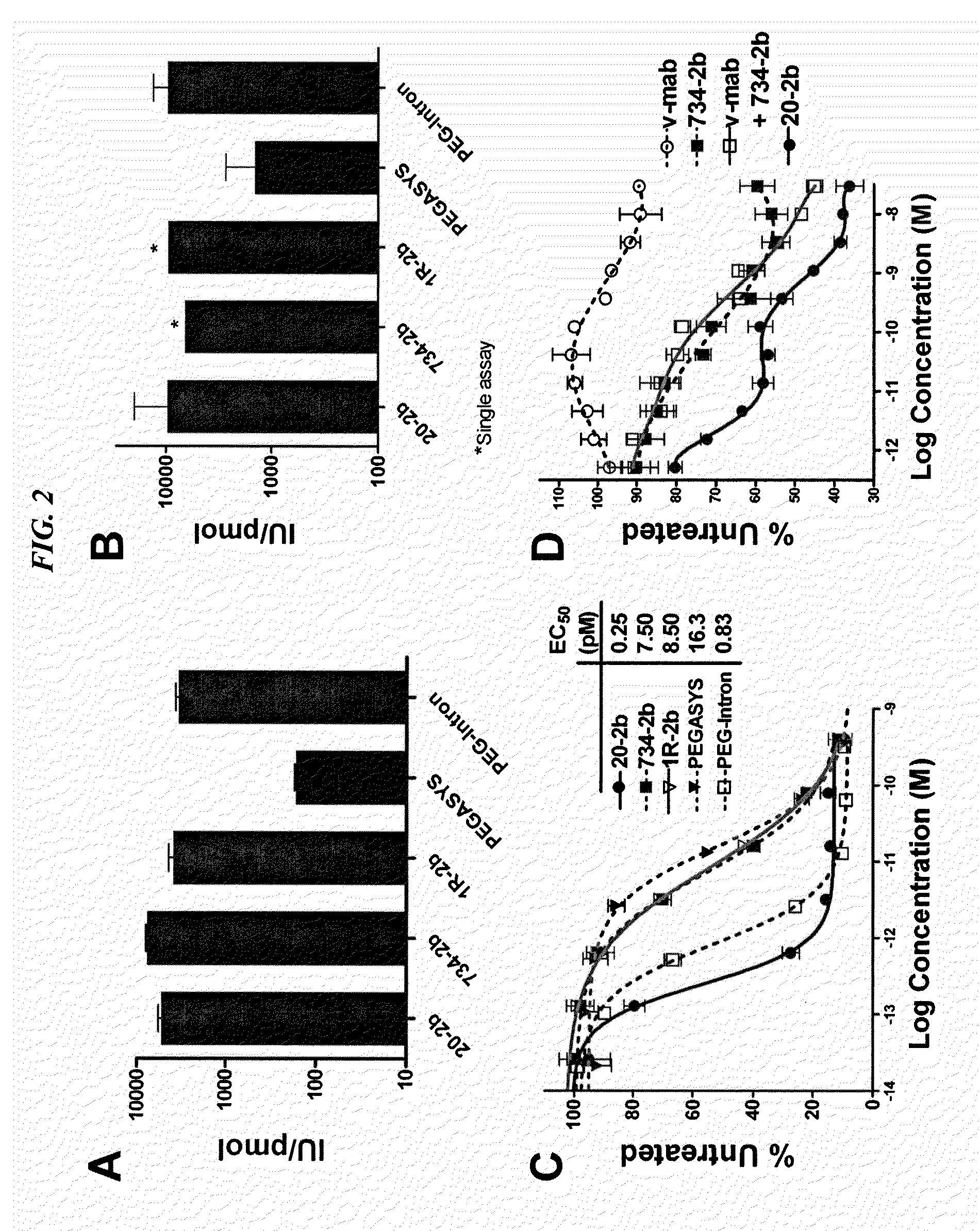
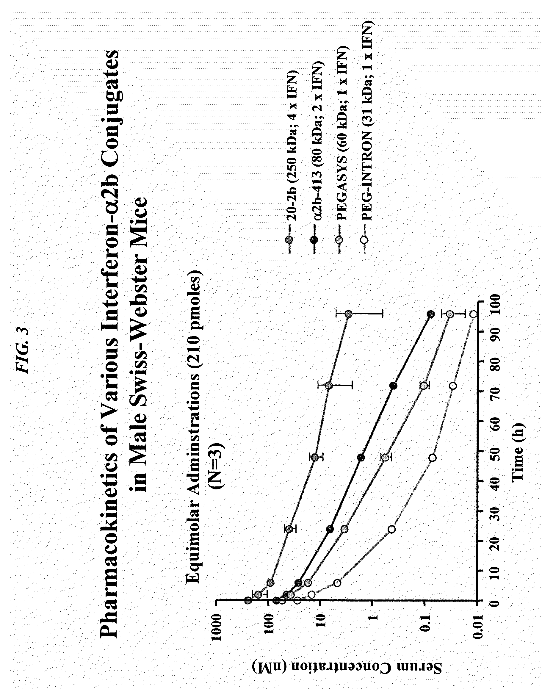
Image

Examples
example 1
CH3-AD2-IgG Expression Vectors
[0157]The pdHL2 mammalian expression vector has been used for the expression of recombinant IgGs (Qu et al., Methods 2005, 36:84-95). A plasmid shuttle vector was produced to facilitate the conversion of any IgG-pdHL2 vector into a CH3-AD2-IgG-pdHL2 vector. The gene for the Fc (CH2 and CH3 domains) was amplified by PCR using the pdHL2 vector as a template and the following oligonucleotide primers:
[0158]
Fc BglII LeftAGATCTGGCGCACCTGAACTCCTG(SEQ ID NO: 21)Fc Bam-EcoRI RightGAATTCGGATCCTTTACCCGGAGACAGGGAGAG.(SEQ ID NO: 22)
[0159]The amplimer was cloned in the pGemT PCR cloning vector (Promega). The Fc insert fragment was excised from pGemT with Xba I and Bam HI and ligated with AD2-pdHL2 vector that was prepared by digesting h679-Fab-AD2-pdHL2 (Rossi et al., Proc Natl Acad Sci USA 2006, 103:6841-6) with Xba I and Bam HI, to generate the shuttle vector Fc-AD2-pdHL2. To convert IgG-pdHL2 expression vectors to a CH3-AD2-IgG-pdHL2 expression vectors, an 861 by ...
example 2
Production of CH3-AD2-IgG
[0166]Transfection and Selection of Stable CH3-AD2-IgG Secreting Cell Lines
[0167]All cell lines were grown in Hybridoma SFM (Invitrogen, Carlsbad Calif.). CH3-AD2-IgG-pdHL2 vectors (30 μg) were linearized by digestion with Sal I restriction endonuclease and transfected into Sp2 / 0-Ag14 (2.8×10⁶ cells) by electroporation (450 volts, 25 μF). The pdHL2 vector contains the gene for dihydrofolate reductase allowing clonal selection as well as gene amplification with methotrexate (MTX).
[0168]Following transfection, the cells were plated in 96-well plates and transgenic clones were selected in media containing 0.2 μM MTX. Clones were screened for CH3-AD2-IgG productivity by a sandwich ELISA using 96-well microtitre plates coated with specific anti-idiotype MAbs. Conditioned media from the putative clones were transferred to the micro-plate wells and detection of the fusion protein was accomplished with horseradish peroxidase-conjugated goat anti-human IgG F(ab′)2 (J...
example 3
Generation of DDD-module Based on Interferon (IFN)-α2b
[0171]Construction of IFN-α2b-DDD2-pdHL2 for Expression in Mammalian Cells
[0172]The cDNA sequence for IFN-α2b was amplified by PCR resulting in sequences comprising the following features, in which XbaI and BamHI are restriction sites, the signal peptide is native to IFN-α2b, and 6 His is a hexahistidine tag: XbaI - - - Signal peptide - - - IFNα2b - - - 6 His - - - BamHI (6 His disclosed as SEQ ID NO: 28). The resulting secreted protein consisted of IFN-α2b fused at its C-terminus to a polypeptide of the following sequence:
[0173]
(SEQ ID NO: 23)KSHHHHHHGSGGGGSGGGCGHIQIPPGLTELLQGYTVEVLRQQPPDLVEFAVEYFTRLREARA.
[0174]PCR amplification was accomplished using a full length human IFNα2b cDNA clone (Invitrogen Ultimate ORF human clone cat # HORFO1Clone ID IOH35221) as a template and the following oligonucleotides as primers:
[0175]
IFNA2 Xba I Left(SEQ ID NO: 24)TCTAGACACAGGACCTCATCATGGCCTTGACCTTTGCTTTACTGGIFNA2 BamHI right(SEQ ID NO: 25)GG...
PUM
| Property | Measurement | Unit |
|---|---|---|
| concentration | aaaaa | aaaaa |
| median survival time | aaaaa | aaaaa |
| MW | aaaaa | aaaaa |
Abstract
Description
Claims
Application Information