System and method for prostate biopsy

a prostate and system technology, applied in the field of prostate biopsy, can solve the problems of affecting the patient's health, affecting the patient's overall health, so as to facilitate planning and facilitate interpretation

Inactive Publication Date: 2010-07-08
EIGEN INC
View PDF4 Cites 32 Cited by
  • Summary
  • Abstract
  • Description
  • Claims
  • Application Information

AI Technical Summary

Benefits of technology

[0024]The systems and methods of the inventions may, in various aspects, also include a subsystem for computing distance from prostate boundaries to facilitate planning. In this subsystem, the system computes and displays the distance of a planned location for sampling a site or placement of radioactive seeds or applicator. Additionally, the system and methods may compute and display iso-surfaces at user-selected distances from prostate surface so as to assist the user in planning and during the procedure. There may be a number of iso-surfaces at user-selected distances from prostate surface to assist the user in planning and during the procedure. For some biopsy procedures, user may want to sample only points within a certain distance from the boundaries of the prostate and a colored iso-surface inside the surface representing the prostate boundaries allows them to do this. In addition, the systems and methods may compute and display the iso-surfaces overlaid on the grayscale image such that a combination of intensity information and boundary information can be utilized in planning a procedure. The computation of distances in 3-D makes it easier for user to interpret 2-D image slices typically seen during planning.
[0025]The systems and methods of the inventions may, in various aspects, also have a subsystem to compute a plan automatically using a user-selected uniform spacing for a procedure, based on prostate boundaries. This makes it easier for procedures where user has to plan uniformly over the prostate for either a biopsy or treatment procedure.
[0026]The systems and methods of the inventions, in various aspects, may also have a subsystem to compute a plan automatically using a user-selected uniform spacing for a procedure, based on prostate boundaries. This makes it easier for procedures where user has to plan uniformly over the prostate for either a biopsy or treatment procedure. In addition, the automatic loading of such a plan also takes into consideration the regions where placing of points is undesirable by avoiding locations close to structures such as urethra and nerve bundles, if such an information is available.
[0027]The systems and methods of the inventions may, in various aspects, also contain a subsystem for correcting any image artifacts caused by misalignment of an ultrasound transducer with respect to the tracking device. The tracking device assumes a known frame of reference with respect to a transducer and as the transducer is manipulated, the tracking device computes the correspondence of the live video frame with the frame of reference of 3-D image. Any misalignment causes artifacts in a reconstructed 3-D image as well as in finding the correspondence between a live 2-D video frame and the 3-D frame of reference. The subsystem for correction image artifacts caused by misalignment of an ultrasound transducer may include applying a known (previously computed or measured) transformation to the transducer frame of reference throughout the procedure. This keeps the correspondence from transducer to the 3-D frame of reference updated throughout the procedure. The setup for computing the transformation may include an ultrasound phantom with landmarks (or beads) placed at known relative locations. The known relative locations in the phantom are used for alignment of the tracking device for the selected transducer. The setup for computing the transformation may also include alignment software which makes it easy for user to scan the ultrasound phantom and computes the transformation automatically after the user follows the directions for scanning the ultrasound phantom.

Problems solved by technology

Such saturation biopsy often causes considerable discomfort to the patient.
Further, smaller organs such a prostate, which may only be 4 cm in diameter, may be damaged by sampling at numerous biopsy sites required for a saturation biopsy.
This becomes overwhelming when a large number of sites are used.
When a patient returns for multiple repeat visits the amount of data and complexity is increased proportionally.

Method used


Image

  • System and method for prostate biopsy
  • System and method for prostate biopsy
  • System and method for prostate biopsy

Examples


Embodiment Construction

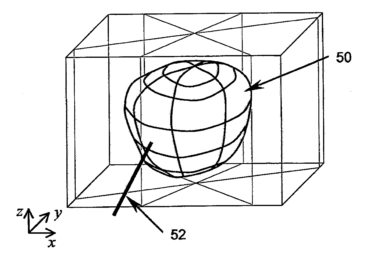
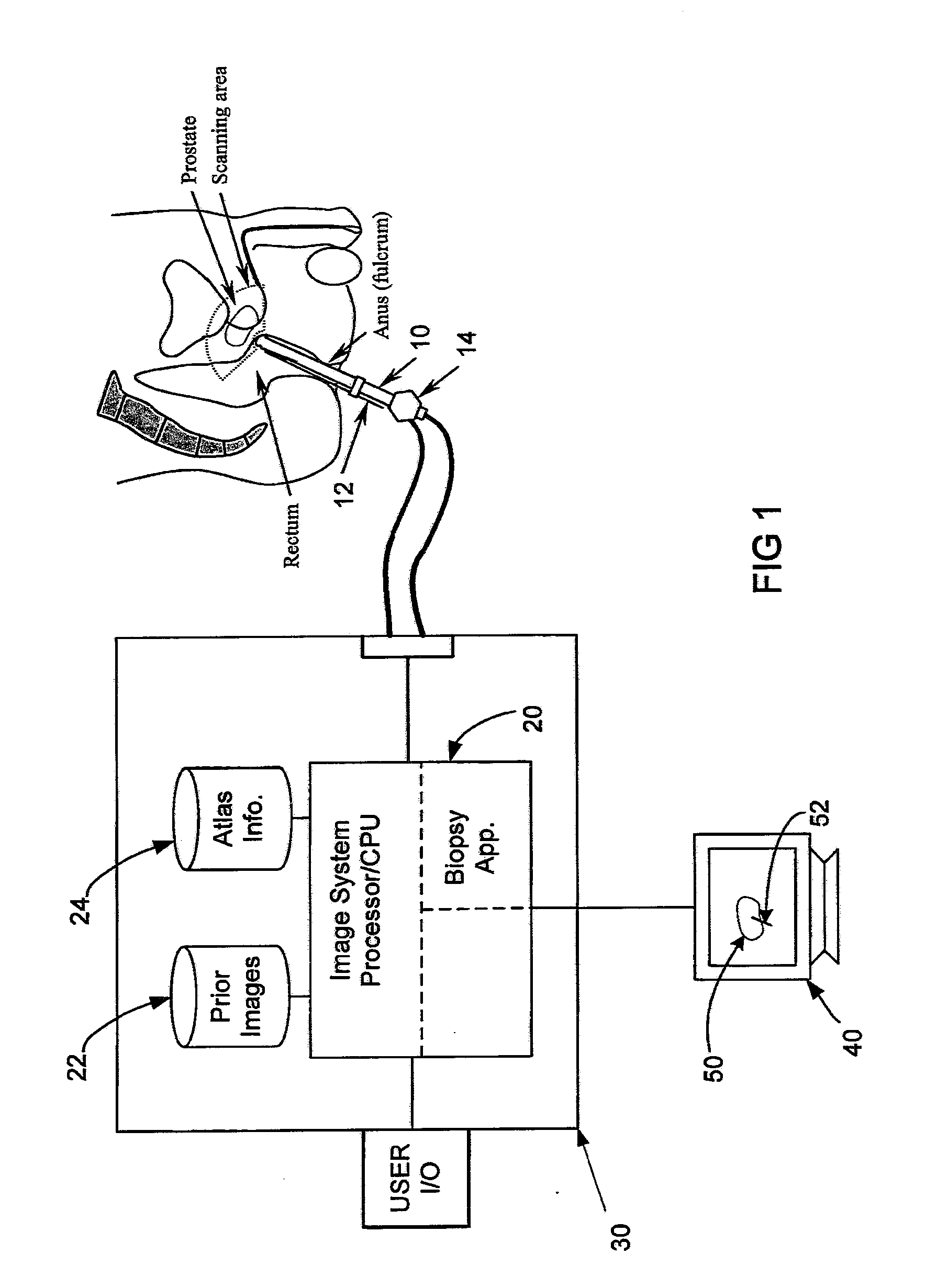
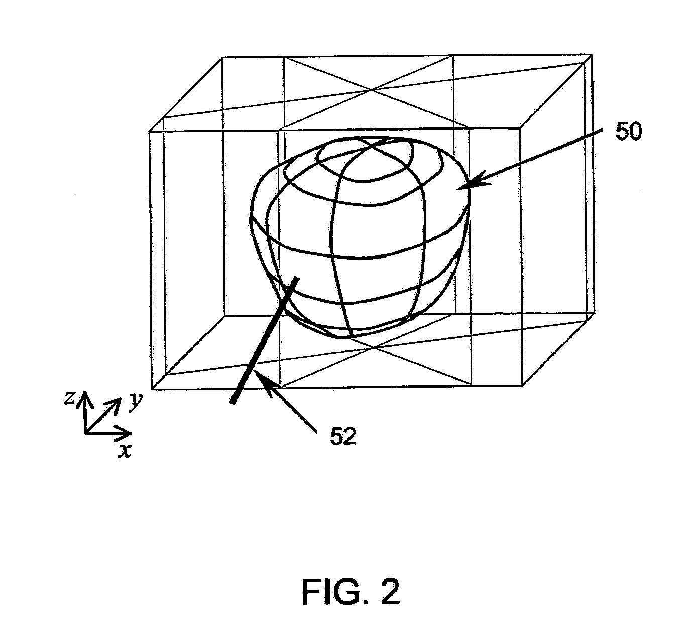
[0044]Reference will now be made to the accompanying drawings, which assist in illustrating the various pertinent features of the various novel aspects of the present disclosure. Although the invention is described primarily with respect to an ultrasound imaging embodiment, the invention is applicable to a broad range of imaging modalities and biopsy techniques, including MRI, CT, and PET, which are applicable to organs and / or internal body parts of humans and animals. In this regard, the following description is presented for purposes of illustration and description. Furthermore, the description is not intended to limit the invention to the form disclosed herein. Consequently, variations and modifications commensurate with the following teachings, and skill and knowledge of the relevant art, are within the scope of the present invention.

[0045]Initially, an exemplary embodiment of the invention will be described in relation to performing prostate biopsy using transrectal ultrasound ...


PUM


Abstract

The invention presents tools to improve a 3-D image aided biopsy or treatment procedure for prostate gland by providing additional functionality and additional visual cues on an output image of the prostate, which may be generated substantially in real-time. The tools include i) the identification of various parts of prostate to classify as per regular classification in pathological reports, ii) Computing and displaying the insertion depth of needle with respect to a selected target point during the procedure, iii) Computing and displaying the distance from needle tip to prostate surface following a procedure and, iv) Calibration for misalignment of a 2-D imaging transducer when used under tracked motion for a procedure.

Description

CROSS-REFERENCE TO RELATED APPLICATION[0001]This application claims priority under 35 U.S.C. 119 to U.S. Provisional Application No. 61 / 113,479, entitled: “Improved System and Method for Prostate Biopsy,” filed on Nov. 11, 2008, the contents of which is incorporated herein as if set forth in full.BACKGROUND[0002]When cancer is suspected on an organ such as a breast, prostate, kidney or liver, a common diagnostic tool is to take biopsies of the organ tissue. A cancer may only be present in a very small portion of the organ in the early stages of its growth. However, this generally represents the stage when it is most desirable to detect the cancer. Cancer may also be uncorrelated and therefore may be present in a very small portion of the organ but at multiple places. Therefore the decision of where in the organ to take a biopsy will have a significant impact on successful early detection of a cancer as well as increasing the confidence of a negative result.[0003]Maximum confidence i...

Claims


Application Information

Patent Timeline
Patent Type & Authority Applications(United States)
IPC IPC(8): G06K9/00
CPCA61B10/0241A61B19/5244A61B2019/5278G06T7/0028G06T2207/30084G06T2207/10132G06T2207/30056G06T2207/30068G06T2207/30081G06T2207/10072A61B2090/3782A61B34/20G06T7/33
Inventor KUMAR, DINESHSURI, JASJIT S.
Owner EIGEN INC
Why Eureka
Try Eureka