
Radiographic film positioning device and a process for obtaining radiographic images

Inactive Publication Date: 2010-08-05
MACHADO ASBEL RODRIGUES +2
View PDF12 Cites 10 Cited by
  • Summary
  • Abstract
  • Description
  • Claims
  • Application Information

AI Technical Summary

Benefits of technology

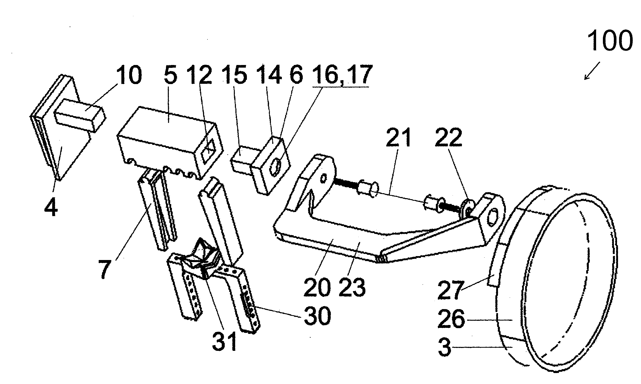
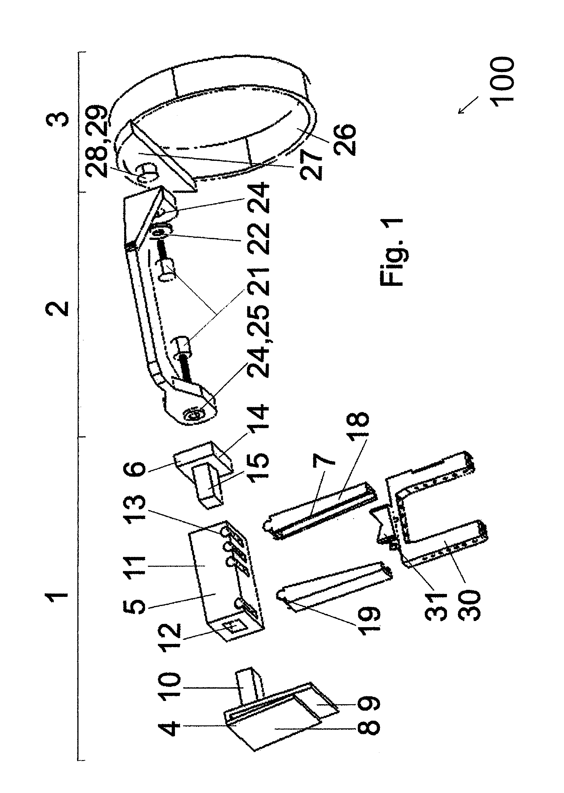
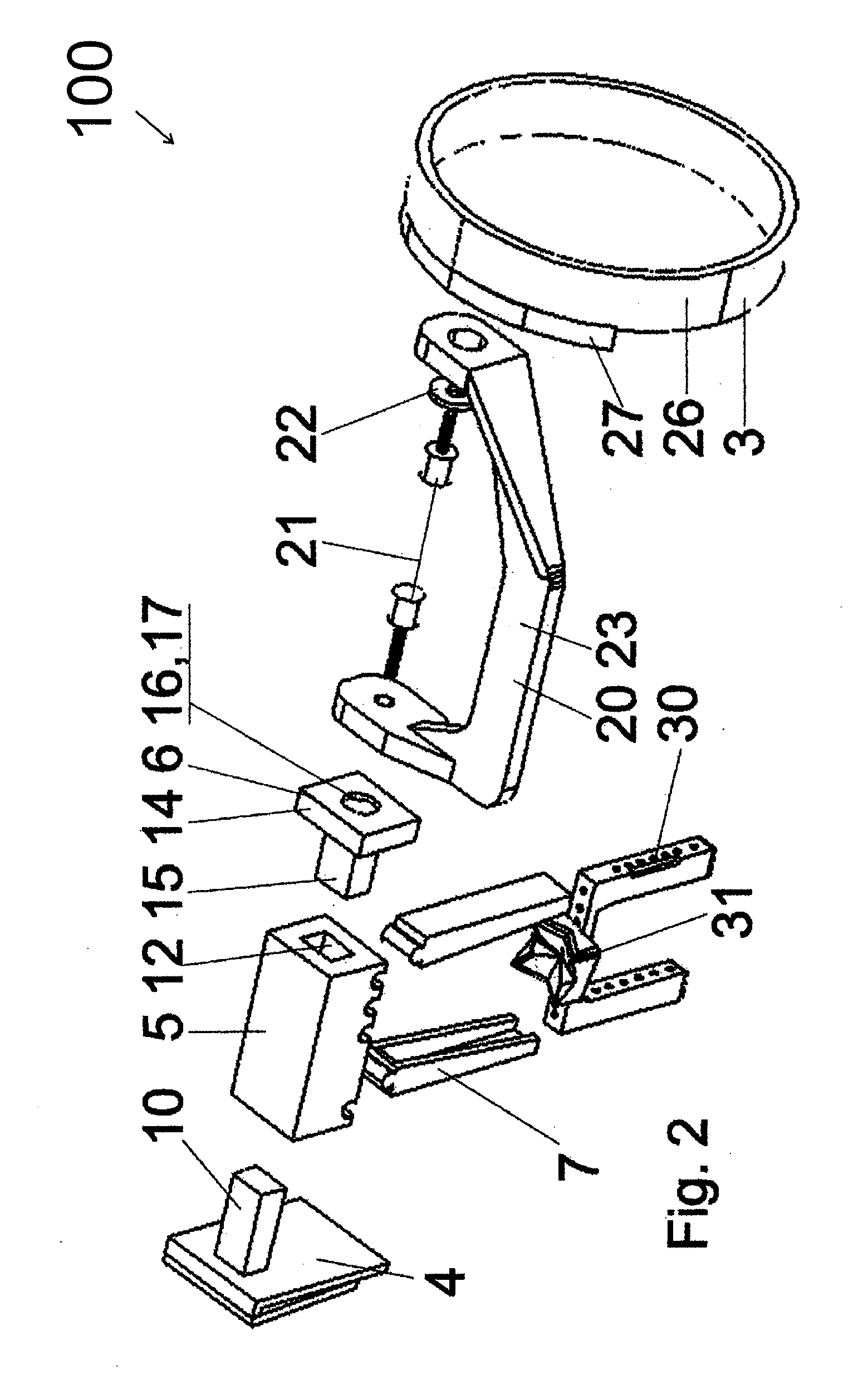
[0041]An objective of the present invention is to provide a radiographic film positioning device, mainly for performing periapical radiographic images, which enables and guarantees great accuracy in parallelism between the radiographic film and the border of the alveolar bone of the patient, thus guaranteeing the obtainment of radiographic images without deformations / distortions due to wrong alignment of the film with respect to the bone.
[0043]A further objective of the present invention is to provide a process for obtaining radiographic images (preferably, but not compulsorily designed for obtaining periapical radiographic images), without such images presenting deformations / distortions due to wrong alignment of the film with respect to the bone.
[0045]Finally, a further objective of the present invention is to provide a process for obtaining radiographic images that provides rigid fixation of the positioner with a radiographic support, so that the image obtained can be perfectly correlated in space with the result of a transgingival probing.BRIEF DESCRIPTION OF THE INVENTION

Problems solved by technology

The radiographic positioners used at present enable the positioning of the film inside the patient's mouth, but do not guarantee parallelism between the film and the border of the patient's alveolar bone.
As a result, the images obtained almost invariably exhibit deformations / distortions due to wrong alignment of the film with respect to the bone, and to not exhibit geometrical and dimensional fidelity.
The procedure of perforating the bone portion of the patient should be carefully studied, since making the perforation at an inadequate site may impair the result of the implantation, in both the esthetic and sometimes the functional aspects, if the fixation becomes difficult due to the incorrect position of the orifice.
In the case of an implant applied to the lower alveolar bone (mandible), there is the aggravating factor of the presence of nerve tissue (lower alveolar nerve) in an inner cavity that goes through the bone, for which reason this situation needs to be considered unfailingly at the time of carrying out the implantation procedure.
If the nerve is reached and damaged, this may result in partial paralysis of the patient's face, becoming permanent sequelae in most cases.
However, if the first orifice was already correctly positioned, then the final orifice of the implant may be impaired.
And, since radiography produces a two-dimensional (height and width) image, which does not enable one to view the third dimension, that is to say, the thickness of the bone border, the professional resorts to a gingival probing examination.
However, the efficiency of all this procedure is impaired because, in spite of the result of the gingival probing, irreparable distortions or deformations of the radiographic images still exist, which are due to limitations in the present radiographic positioners, which are not able to guarantee parallelism between the radiographic film and the border of the patient's alveolar bone, unless by a stoke of luck.
As serious drawbacks, the device disclosed ion document BRMU 6400302-7 does not provide rigid fixation and does not guarantee, with sufficient precision, perfect positioning and parallelism of the film with the object being X-rayed.
Such failure results from the fact that fixation in a given position is guaranteed by occlusion, so that, when the patients bites, displacement and inclination of the positioner may occur (and usually occurs), depending on the anatomy of the teeth and on the intensity of the bite.
The device disclosed in that document, in spite of presenting rigid fixation by means of the resin, does not provide means for guaranteeing parallelism, because the resin only fixes the device in the position in which it is, nor does it teach means for guaranteeing that this positioning is correct, keeping the radiographic film parallel to the bone.
As another drawback, this is a quite toilsome procedure, since the making of the bite guide with resin is always individualized, specifically intended only for that patient, which requires more time and makes the process expensive.
This device is specific for application in endodontical treatment and is not effective for making radiographic shots with a view to making implants, because, since it does not have rigid fixation, it cannot guarantee with sufficient accuracy the perfect positioning and parallelism of the film with the object being X-rayed (bone).
This failure results from the fact that the fixation in a given position is guaranteed by occlusion, so that, when the patient bites, displacement and inclination of the positioner may occur depending on the anatomy of the teeth and on the intensity of the bite.
So far, no process for obtaining radiographic images (preferably, but not compulsorily designed for obtaining periapical radiographic images) had been developed, without such images presenting deformations / distortions due to wrong alignment of the film with respect to the bone.

Method used


Image

  • Radiographic film positioning device and a process for obtaining radiographic images
  • Radiographic film positioning device and a process for obtaining radiographic images
  • Radiographic film positioning device and a process for obtaining radiographic images

Examples


Embodiment Construction

[0057]A dental implant is usually employed to recover the appearance of the mouth of a patient who has definitively lost one of more teeth.

[0058]As it is known, in addition to enabling mastication and chewing of foods in small portions (capable of going through the esophagus), the teeth have a number of important actuations, among which the esthetic appearance which they impart to people and still the influence which they exert on certain phonemes of speech, the pronunciation of which in the absence of the teeth are impaired. Therefore, the presence of teeth in the mouth is very important, from both the esthetic and functional point of view.

[0059]Typically, the procedure followed for a dental implant comprises using a synthetic tooth (technically referred to as prosthetic crown), which should be positioned at the site of the missing original tooth, for the purpose of restoring the capability of chewing and other properties attributed to the tooth.

[0060]A crown of tooth is fixed to a...


PUM


Abstract

One describes a radiographic film positioning device, particularly de-signed for use in dentistry, provided with a first assembly for positioning at least one radiographic film that comprises at least one association means for association with a radiographic support, positionable substantially perpendicular to a bone portion of the patient.

Description

[0001]The present invention relates to a radiographic film positioning device, particularly, but not compulsorily designed for use in dentistry, which guarantees correct positioning of the film with respect to the alveolar bones of the patient's mouth and, as a result, enables one to obtain precise true radiographic images, without deformations resulting from wrong alignment of the film with respect to the bone.[0002]The present invention has the objective of providing a radiographic film positioning device that enables rigid fixation thereof with a radiographic support, so that the radiographic image produced can be perfectly correlated in space with the contour of the bone relief obtained with a transgingival probing carried out with said support, thus enabling one to determine the ideal values of the position and of the angle of a dental implant which is to be installed in the region being X-rayed.[0003]The accuracy of the radiographic images obtained makes the present positionin...

Claims


Application Information

Patent Timeline
IPC IPC(8): A61B6/14
CPCA61B6/14A61B6/4435A61B6/145A61B6/506A61B6/512A61B6/51
Inventor MACHADO, ASBEL RODRIGUESRANGEL, EDER FERREIRARANGEL, KEULER FERREIRA
Owner MACHADO ASBEL RODRIGUES
Learn More