Automatic outcome analysis using radiological images

a radiological image and automatic technology, applied in the field of radiological analysis, can solve the problem of difficult quantitative evaluation of the outcome of treatment by visual inspection alon

Inactive Publication Date: 2010-08-26
GENERAL ELECTRIC CO
View PDF16 Cites 9 Cited by
  • Summary
  • Abstract
  • Description
  • Claims
  • Application Information

AI Technical Summary

Problems solved by technology

It may be difficult to quantitatively evaluate the outcomes of treatment by visual inspection alone.

Method used


Image

  • Automatic outcome analysis using radiological images
  • Automatic outcome analysis using radiological images
  • Automatic outcome analysis using radiological images

Examples


Embodiment Construction

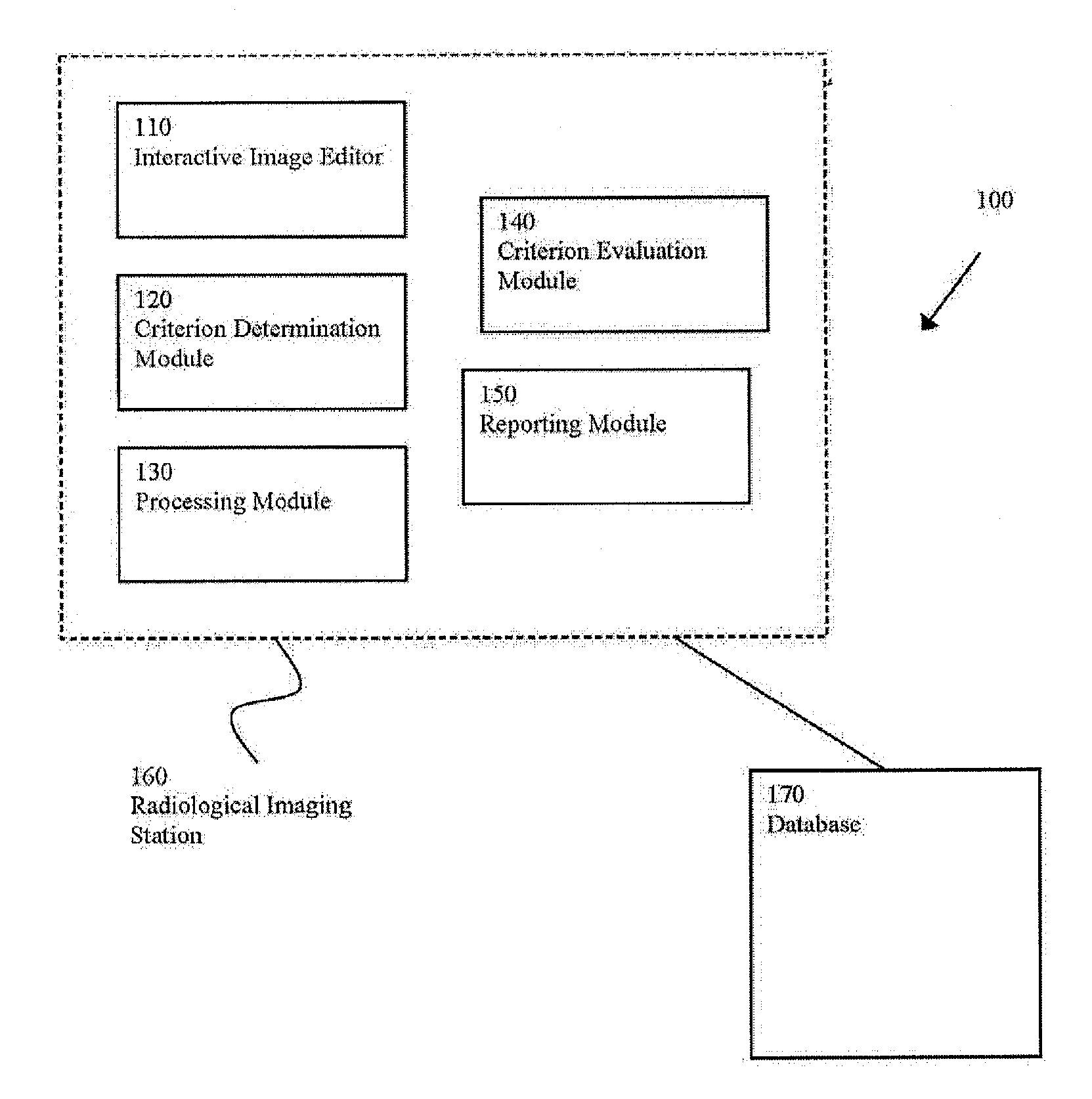
[0012]FIG. 1 shows a block diagram 100 of a system for analyzing a treatment of a patient, in accordance with an embodiment of the present invention. System 100 includes an interactive image editor 110, a criterion determination module 120, a processing module 130, a criterion evaluation module 140, a reporting module 150, a radiological imaging station 160, and a database 170. One or more portions of system 100, such as radiological imaging station 160, may be incorporated into an enterprise system, such as a radiological information system (“RIS”), or picture archiving and communication system (“PACS”). The components may be centrally located, or may be distributed, for example, across a network. The components may be physical and / or logical. The components may be implemented as software or firmware, for example, by a computer readable storage medium having a set of instructions.

[0013]The interactive image editor 110 allows a user, such as a radiologist or other clinician, to inte...


PUM


Abstract

Certain embodiments of the present invention provide a system for analyzing a treatment of a patent including: an interactive image editor for allowing a user to interact with a first set image data corresponding to a pre-treatment radiological study of a patient, the first set of image data including an untreated clinical area of interest, such that the user is capable of interacting with the first set of image data to form a target outcome image for the clinical area of interest; a criterion determination module for allowing the user to determine a target outcome criterion; processing module for performing a comparison by comparing the first set of image data with a second set of image data corresponding to a post-treatment radiological study of the patient, the second set of image data including a treated clinical area of interest; and a criterion evaluation module for automatically evaluating the comparison to determine a post-treatment effect. In an embodiment, the criterion evaluation module is further for evaluating automatically a treatment effectiveness evaluation based on an extent to which the post-treatment effect satisfies the target outcome criterion.

Description

FEDERALLY SPONSORED RESEARCH OR DEVELOPMENT[0001][Not Applicable]MICROFICHE / COPYRIGHT REFERENCE[0002][Not Applicable]BACKGROUND OF THE INVENTION[0003]Embodiments of the present invention relate generally to analysis of radiological studies. Specifically, embodiments of the present invention relate to providing quantitative and visual analysis of pre and post treatment radiological studies.[0004]Radiology imaging can be used to image a patient and a clinical area of interest before, and after a procedure. In order to determine whether the procedure is successful, the clinician (e.g., radiologist or treating physician) may visually compare the pre and post-treatment images. It may be difficult to quantitatively evaluate the outcomes of treatment by visual inspection alone. Clinicians may have desired outcomes, and they may be described in text or as notes on the pre-treatment radiological study. In addition to visual inspection, it may be helpful to provide a clinician with a quantita...

Claims


Application Information

Patent Timeline
Patent Type & Authority Applications(United States)
IPC IPC(8): G06K9/00G06K9/54G16H20/40G16H30/20G16H30/40
CPCA61B19/50A61B2019/504G06F19/321G06T11/60G06F19/3437G06F19/3487G06F19/3425A61B2034/104A61B34/10G16H80/00G16H50/50G16H15/00G16H30/20G16H20/40G16H30/40
Inventor WOO, YIJIN
Owner GENERAL ELECTRIC CO
Why Eureka
Try Eureka