System and method for non-invasive monitoring of cerebral tissue hemodynamics

a non-invasive, cerebral tissue technology, applied in the field of human vital functions monitoring, can solve the problems of neurological damage, severe, irreversible, non-invasive methods such as pulse oximtery cannot be used to obtain cerebral venous blood oxygenation, and it is difficult for light to penetrate through the skull and reach the cerebral tissues

Active Publication Date: 2010-09-02
MESPERE LIFESCI
View PDF8 Cites 12 Cited by
  • Summary
  • Abstract
  • Description
  • Claims
  • Application Information

AI Technical Summary

Benefits of technology

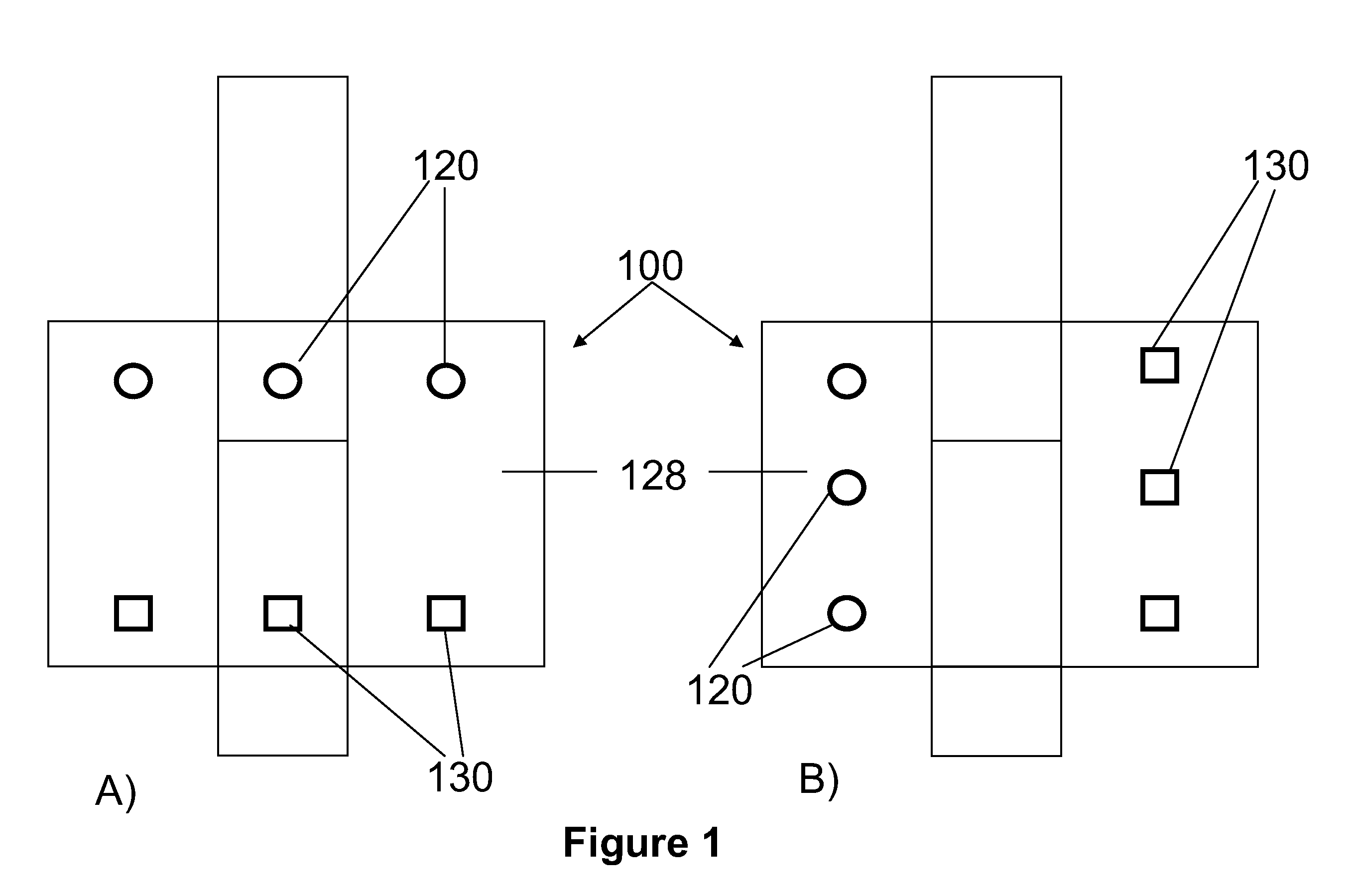
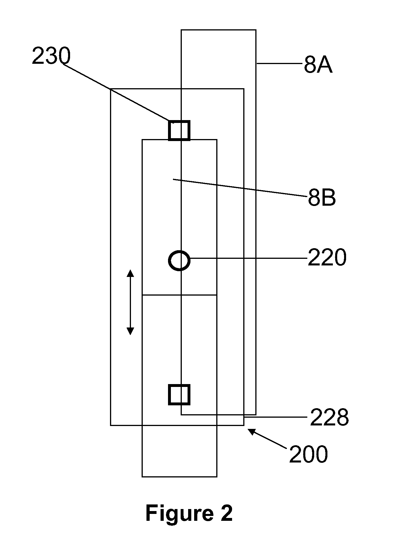
[0006]The present invention provides a novel method of determining cerebral tissue oxygenation, as well as a system useful for such a determination. The method includes optically obtaining an output signal of internal jugular venous blood at at least two different w...

Problems solved by technology

Insufficient cerebral tissue oxygen consumption over a few minutes can lead to irreversible, severe, neurological damage.
Non-invasive methods such as pulse oximtery cannot be used to obtain cerebral venous blood oxygenation because, unlike arterial vessels, venous blood vessels are passive and without pulsation...

Method used


Image

  • System and method for non-invasive monitoring of cerebral tissue hemodynamics
  • System and method for non-invasive monitoring of cerebral tissue hemodynamics
  • System and method for non-invasive monitoring of cerebral tissue hemodynamics

Examples


example 1

Measurement of Cerebral Oxygenation in a Patient

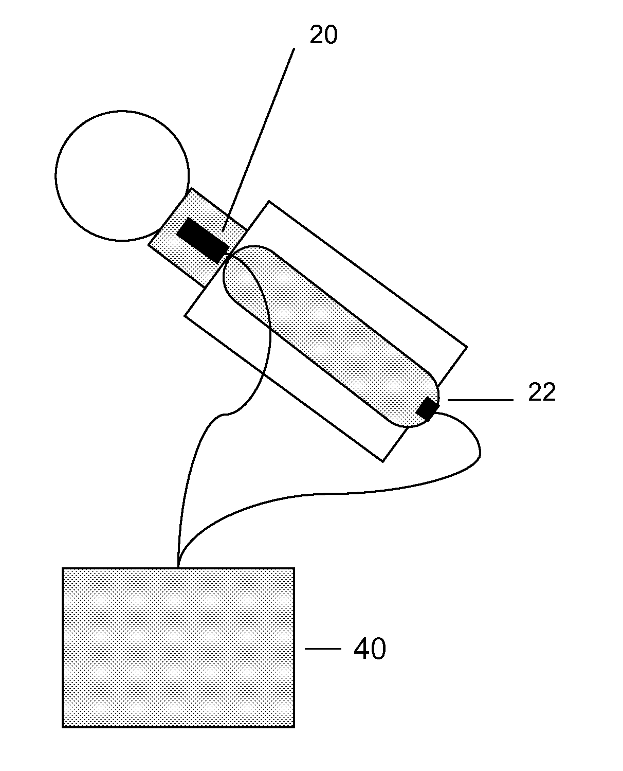
[0061]The cerebral tissue oxygenation of a human subject was obtained using a system as shown in FIG. 6. The patient lay on a chair at about a 30 degree recline. The first module of the system, e.g. sensor patch, was placed on the neck of the patient at a site over the internal jugular vein. The second module (oximeter sensor patch) was placed on the index finger tip of the patient. While the patient maintained normal breathing, jugular venous pulse (output signal) was obtained from the first module and cardiac arterial pulse (output signal) was obtained from the second module. The signals were received by the signal processing module, processed as described above, to yield a jugular venous waveform (FIG. 7A), a cardiac arterial waveform (FIG. 7B) and a cerebral venous waveform (FIG. 7C). The amplitude of the detected signals is represented along the y-axis while the x-axis represents time. The signal processing module utilized this si...


PUM


Abstract

A method and system are provided which are useful for the non-invasive determination and monitoring of cerebral tissue oxygenation. The method comprises the steps of generating at least first and second jugular venous output signals against time based on the reflection of at least first and second wavelengths of light, respectively, from an external tissue site on the patient in the proximity of the internal jugular vein; obtaining corresponding first and second cardiac arterial output signals for the first and second wavelengths of light, respectively, from the patient, and separating the first and second cardiac arterial output signals from the first and second jugular venous output signals, respectively, to generate first and second cerebral venous output signals; and determining cerebral tissue oxygenation based on the first and second cerebral venous output signals. A system useful to monitor cerebral tissue oxygenation may comprise a first module for optically generating at least first and second jugular venous output signals against time at at least first and second wavelengths of light, respectively, from the patient; a second module for generating first and second cardiac arterial output signals at the first and second wavelengths of light, respectively, from the patient; and a signal processing means adapted to separate the first and second cardiac arterial output signals from the first and second jugular venous output signals, respectively, to yield first and second cerebral venous output signals, for the determination of cerebral tissue oxygenation.

Description

[0001]This application claims the benefit of U.S. Provisional Application No. 61 / 202,441, filed Feb. 27, 2009, and incorporates such provisional application in its entirety by reference.FIELD OF THE INVENTION[0002]The present invention is related to techniques for monitoring vital functions of the human body, including cerebral tissue hemodynamics. It relates, in particular, to an optical method and system for the non-invasive and continuous monitoring of cerebral parameters such as cerebral tissue oxygenation.BACKGROUND OF THE INVENTION[0003]Monitoring cerebral tissue oxygenation or oxygen consumption is critical for the management of patients with traumatic brain injury and patients undergoing cardiac by-pass surgeries. Insufficient cerebral tissue oxygen consumption over a few minutes can lead to irreversible, severe, neurological damage. Cerebral oxygen consumption can be estimated through the differences between arterial and cerebral venous blood oxygen saturation. Pulse Oximet...

Claims


Application Information

Patent Timeline
IPC IPC(8): A61B5/1455
CPCA61B5/6833A61B5/14553A61B5/4064
Inventor CHENG, XUEFENG
Owner MESPERE LIFESCI
Why Eureka
Try Eureka