Method and apparatus for field map estimation

a field map and estimation method technology, applied in the field of magnetic resonance imaging (mri), can solve problems such as degrading the diagnostic value of mr images

Active Publication Date: 2010-11-11
THE BOARD OF TRUSTEES OF THE LELAND STANFORD JUNIOR UNIV
View PDF14 Cites 20 Cited by
  • Summary
  • Abstract
  • Description
  • Claims
  • Application Information

AI Technical Summary

Benefits of technology

[0008]In an embodiment of the invention, the method for estimating values of a field map includes fixing a field map value for a pixel to a most likely field map value if the pixel satisfies a smoothness constraint with its neighboring pixels. In another embodiment of the invention, the method for estimating values of a field map includes fixing a field map value for a pixel to a value adjacent to a most likely field map value if the pixel does not satisfy a smoothness constraint with its neighboring pixels. In some embodiments of the invention, the smoothness constraint minimizes a number of inconsistent pairs of field map values for neighboring pixels in the field map.

Problems solved by technology

Unless suppressed, bright fat signals may obscure underlying pathology, degrading the diagnostic value of MR images.

Method used


Image

  • Method and apparatus for field map estimation
  • Method and apparatus for field map estimation
  • Method and apparatus for field map estimation

Examples


Embodiment Construction

[0022]Compared to fat suppression methods based on spectrally-selective excitation, water-fat separation methods based on chemical-shift induced phase (also referred to as phase-sensitive water-fat separation methods), such as the 3-point Dixon method described by G. H. Glover, “Multipoint Dixon Technique for Water and Fat Proton and Susceptibility Imaging,” (J. Magn. Reson. Imaging., 1991, vol. 1, no. 5, pp. 521-530) and the IDEAL method described in U.S. Pat. No. 7,176,683 B2 and incorporated by reference herein, show better immunity to field inhomogeneities. Phase-sensitive water-fat separation estimates field inhomogeneities from multiple MR images acquired at different echo times, which is a simple case of spectroscopic imaging. The most common acquisition schemes fall into two categories, two-point and three-point acquisitions, according to the number of the acquired MR images. Three-point acquisition with flexible echo times has shown success in many clinical applications and...


PUM


Abstract

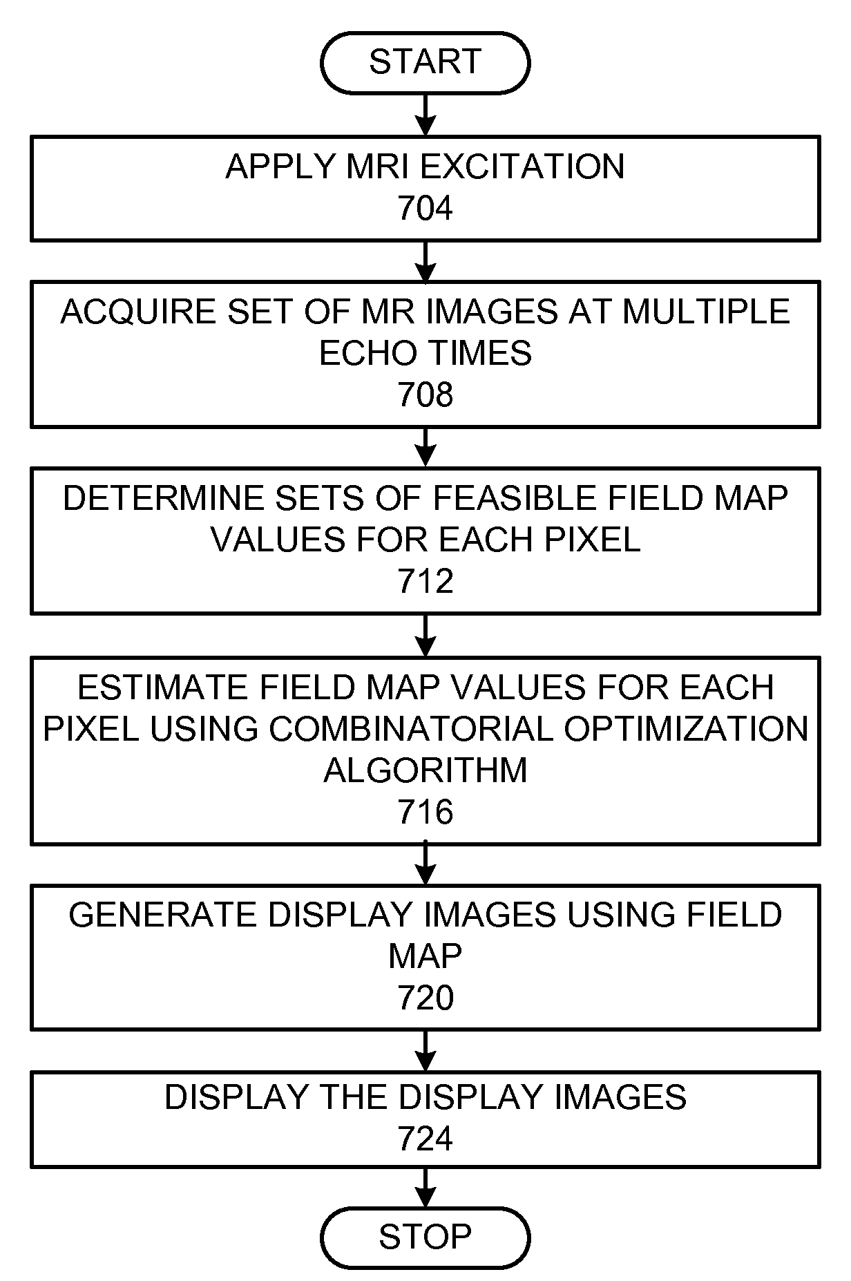
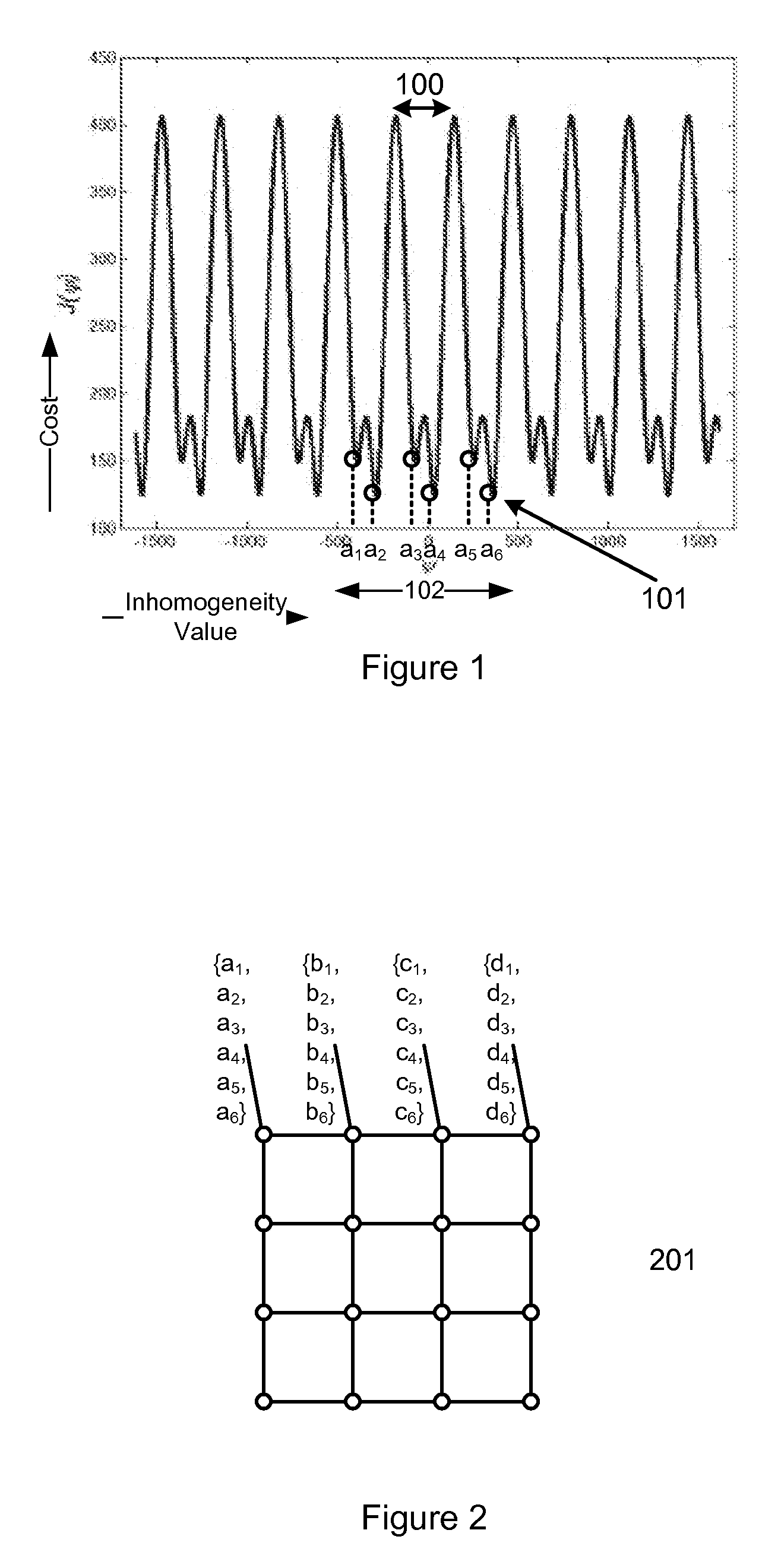
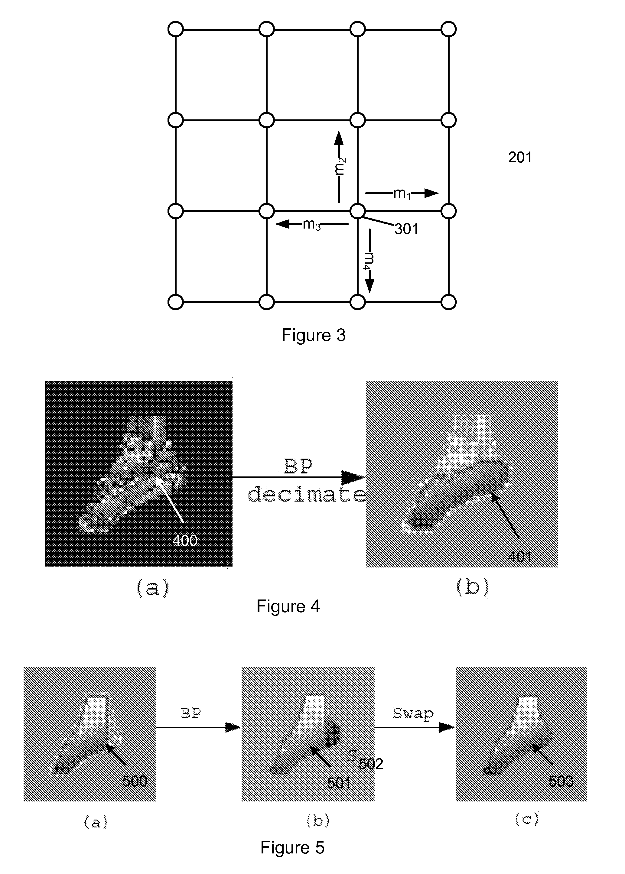
A method for estimating values of a field map to generate a magnetic resonance display image with species separation is provided. A set of MR images is acquired based on an applied magnetic resonance excitation. A set of feasible field map values for each pixel in a field map are determined from the set of MR images. Estimated values of the field map for each pixel are chosen from the set of feasible field map values using a combinatorial optimization algorithm that includes a smoothness constraint. The combinatorial optimization algorithm includes iteratively communicating, between neighboring pixels in the field map, sum-product belief messages that include likelihoods for feasible field map values. Field map values are fixed to most likely field map values if the pixel satisfies the smoothness constraint with its neighboring pixels. A magnetic resonance display image with species separation is generated using the estimated field map.

Description

GOVERNMENT RIGHTS[0001]The U.S. government has rights in the disclosed invention pursuant to NIH grants to Stanford University including NIH-EB002524-01, NIH-RR009784-11, NIH-HL075803-01.BACKGROUND OF THE INVENTION[0002]This invention relates generally to magnetic resonance imaging (MRI), and more particularly, the invention relates to field map estimation from multi-echo MRI sequences.[0003]Magnetic resonance imaging (MRI) is a non-invasive method for the analysis of materials and is an approach to medical imaging. It does not involve ionizing radiation. In very general terms, nuclear magnetic moments are excited at specific spin precession frequencies which are proportional to the local magnetic field. The radio-frequency signals resulting from the precession of these spins are received using pickup coils. By manipulating the magnetic fields, an array of signals is provided representing different regions of the volume. These are combined to produce a volumetric image of the nuclea...

Claims


Application Information

Patent Timeline
Patent Type & Authority Applications(United States)
IPC IPC(8): G01R33/44
CPCG01R33/243G01R33/4828G01R33/443
Inventor LU, WENMIAOLU, YI
Owner THE BOARD OF TRUSTEES OF THE LELAND STANFORD JUNIOR UNIV
Why Eureka
Try Eureka