
Microscopic imaging techniques

a microscopy and imaging technology, applied in the field of microscopy imaging, can solve the problems of not providing any axial resolution, only having a very poor axial resolution, and not providing an improved 3d resolution in the axial direction

Active Publication Date: 2010-11-11
JACKSON LAB THE +1
View PDF55 Cites 28 Cited by
  • Summary
  • Abstract
  • Description
  • Claims
  • Application Information

AI Technical Summary

Problems solved by technology

However, a problem with current FPALM imaging techniques is that it does not provide improved 3D resolution in the axial direction.
Although FPALM can provide a 3D image of a particle by combining images of multiple 2D planes of a sample, this type of image has only a very poor resolution in the axial direction comparable to conventional microscopy because it fails to provide any axial resolution within any of the combined images.
Thus, FPALM has not yet been extended to 3D imaging.
This is due to the currently used standard widefield or total internal reflection (TIR) microscopy geometries not being suitable to 3D FPALM imaging.
None of the current fluorescence microscopy techniques can enhance resolutions of light microscopy to levels of electron microscopy.

Method used


Image

  • Microscopic imaging techniques
  • Microscopic imaging techniques
  • Microscopic imaging techniques

Examples


Embodiment Construction

[0025]While this invention is susceptible of embodiment in many different forms, there is shown in the drawings and will herein be described in detail preferred embodiments of the invention with the understanding that the present disclosure is to be considered as an exemplification of the principles of the invention and is not intended to limit the broad aspect of the invention to the embodiments illustrated.

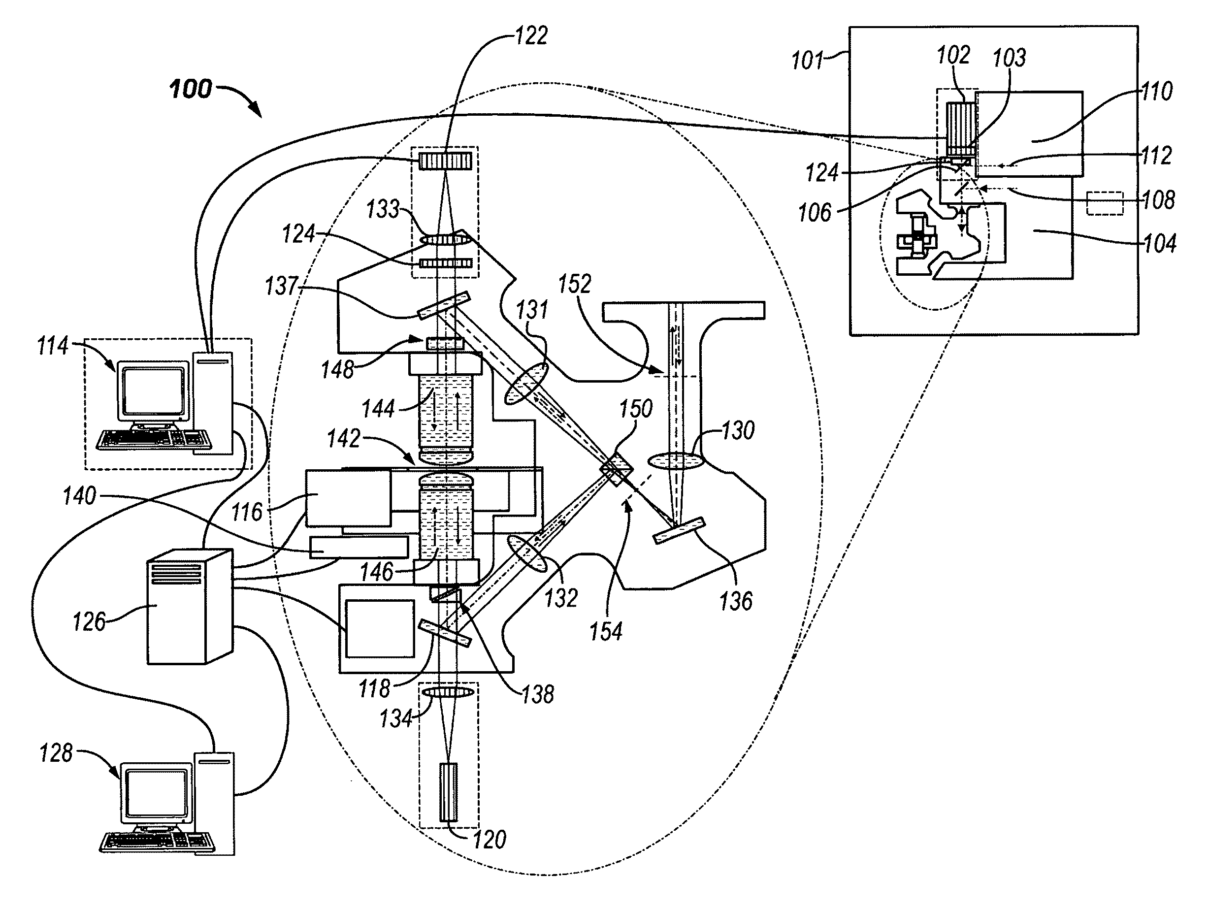
[0026]According to one embodiment, Three-Dimensional Fluorescence Photo Activation Localization Microscopy (“3D FPALM”) methods provide a localization-based resolution of at least 30 nanometers in three-dimensions (“3D”) that may be achieved on a single molecule level. In fact, the 3D FPALM methods can resolve particles on the order of about 20-30 nanometers in the lateral plane (more than 10 times greater than conventional, 4Pi, and I5M microscopy) and on the order of about 10 nanometers in the axial direction (about 60 times greater than conventional microscopy and about 10 ti...


PUM


Abstract

A method of performing 3D photoactivation microscope imaging includes providing a sample having a plurality of probes, each of the plurality of probes including a photo-activatable material. Probes from the plurality of probes are activated to form a sparse subset of probes, the sparse subset of probes having probes that are spatially separated by at least a microscope resolution. The sample is illuminated with a readout light source, and light emitted from activated probes is detected. Based on the light emission detected from the activated probes, localized three-dimensional positions of the activated probes are obtained.

Description

RELATED APPLICATIONS[0001]This application is related to and claims priority to U.S. Provisional Patent Application Ser. No. 60 / 879,833 filed Jan. 11, 2007, titled “Microscopic Imaging Techniques,” which is incorporated herein in its entirety.FIELD OF THE INVENTION[0002]The present invention relates generally to microscopic imaging and, more specifically, to three-dimensional (“3D”) photoactivation localization microscope imaging.BACKGROUND OF THE INVENTION[0003]A conventional light microscope provides a resolution on the order of about 250 nanometers in a lateral two-dimensional (“2D”) plane (i.e., x-y plane) and on the order of about 600 nanometers, or worse, in an axial direction (i.e., z axis). To obtain higher resolution images, other microscopy methods must be utilized.4Pi-Microscopy[0004]A number of techniques have been able to provide a higher resolution in the axial direction to achieve an improved 3D image of structures. For example, 4Pi confocal microscopy and I5M microsc...

Claims


Application Information

Patent Timeline
Patent Type & Authority Applications(United States)
IPC IPC(8): H04N13/02
CPCG02B21/0076G02B21/16G02B27/58G02B21/18G02B21/367
Inventor BEWERSDORF, JOERGMASON, MICHAEL DARINHESS, SAM T.
Owner JACKSON LAB THE
Learn More