Surgical system

a biopsy and surgical technology, applied in the field of biopsy systems and, can solve the problems of increasing the risk of infection and bleeding at the sample site, significant trauma to the breast tissue, and requiring considerable recovery time for the patient,

Active Publication Date: 2011-01-06
SUROS SURGICAL SYST
View PDF105 Cites 48 Cited by
  • Summary
  • Abstract
  • Description
  • Claims
  • Application Information

AI Technical Summary

Benefits of technology

[0017]A surgical device is disclosed that comprises a cutting element, a biopsy line, a motor line, and a vacuum line. The cutting element includes an outer cannula and an inner cannula. The biopsy line is operatively connected to a control console for moving the inner cannula within the outer cannula to cut tissue cores. The motor line is operatively connected to the control console for operating the surgical device. The vacuum line is operatively connected to the inner cannula. A selectively openable tissue filter is operatively connected to the inner cannula, and is also connected to the vacuum line.

Problems solved by technology

This procedure causes significant trauma to the breast tissue, often leaving disfiguring results and requiring considerable recovery time for the patient.
This is often a deterrent to patients receiving the medical care they require.
The open technique, as compared to the percutaneous method, presents increased risk of infection and bleeding at the sample site.
In addition, this method does not provide for a pathological assessment of the tissue, which can provide a more complete assessment of the stage of the cancer, if found.
First, the device would sometimes fail to remove a sample, therefore, requiring additional insertions.
This was generally due to tissue failing to prolapse into the sampling notch.
Secondly, the device had to be inserted and withdrawn to obtain each sample, therefore, requiring several insertions in order to acquire sufficient tissue for pathology.
While the vacuum assisted core biopsy device presented an advancement in the field of biopsy devices, several disadvantages remain with some of the currently marketed devices.
Because the tubes are attached to various components of the biopsy device, all of these tubes (including tubes that do not come into contact with bodily fluids) are disposed of at the conclusion of the procedure, leading to waste and increased expense.
Another issue with current biopsy devices is insuring proper placement of a filter in a tissue collection chamber.
Because the filter is a separate removable piece from the collection chamber, during assembly of the biopsy device it may be inadvertently omitted from the collection chamber.
However, if the filter is not properly placed within the tissue collection chamber, the resected tissue is not properly retained within the filter.
Instead, resected tissue cores may pass through the tissue collection chamber and become lodged in the vacuum line, disabling the biopsy device and prohibiting examination of the cores.
Another issue with prior art devices relates to different modes of operation of the device.
And if a physician attempts to tale tissue cores while the device is set in the lavage mode, the device will not operate, triggering malfunction alarms.
Another issue that may be experienced by users of the biopsy devices is not being able to cock the biopsy device (i.e., preparing the device for firing by retracting the outer cannula) for taking biopsy cores while the device is in the lavage mode.
And if the device is cocked and fired in the lavage mode, the tissue aperture will be open, causing additional trauma to the patient.
Even if the biopsy device is in the cocked position, there still may be an issue with acquiring tissue.
If the remote firing valve is not placed in the correct position, such as placing the remote valve between the cocked the fired positions so as only to partially cock the device, the device will not fully fire during the procedure and tissue may not be fully cut during the biopsy cycles.

Method used


Image

  • Surgical system
  • Surgical system
  • Surgical system

Examples


Embodiment Construction

[0038]Referring now to the drawings, the preferred illustrative embodiments of the present disclosure are shown in detail. Although the drawings represent some embodiments of the present disclosure, the drawings are not necessarily to scale and certain characteristics may be exaggerated to better illustrate and explain the present disclosure. Further, the embodiments set forth herein are not intended to be exhaustive or otherwise limit or restrict the disclosure to the precise forms and configurations disclosed in the following detailed description.

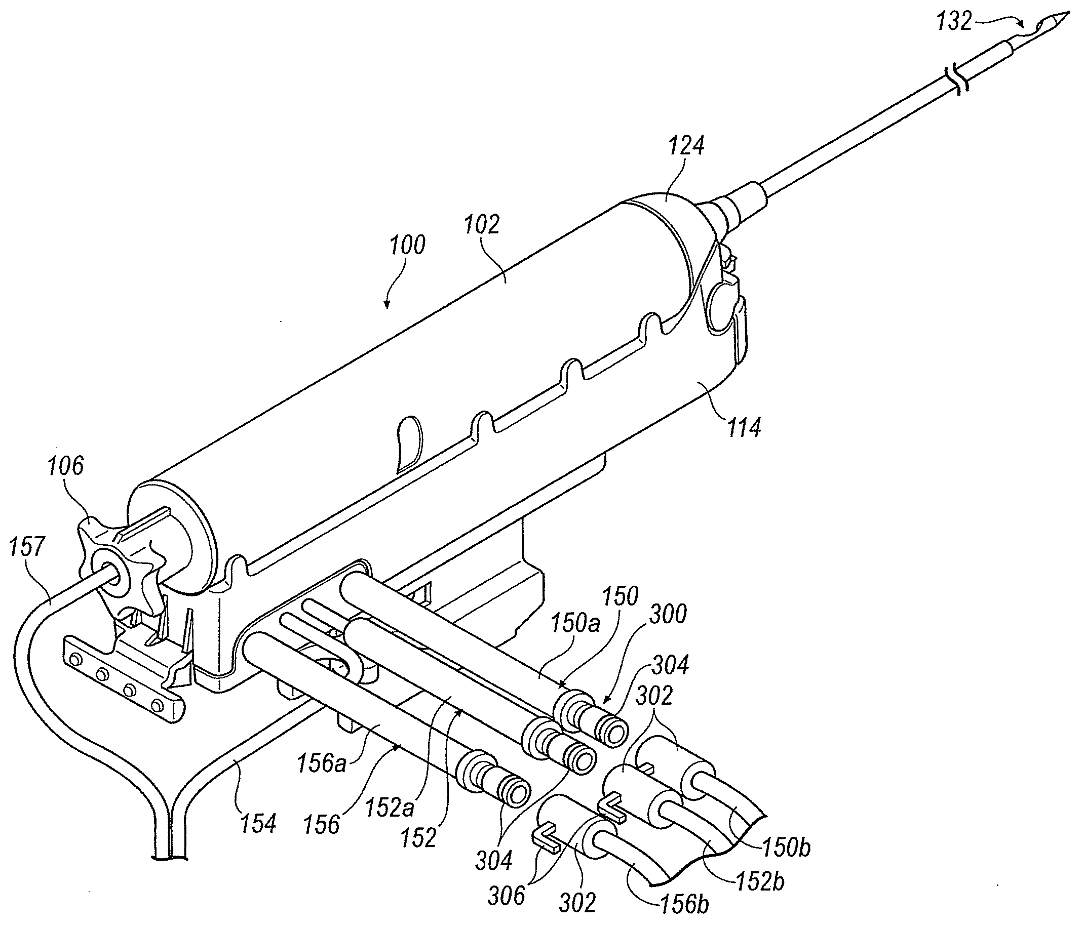
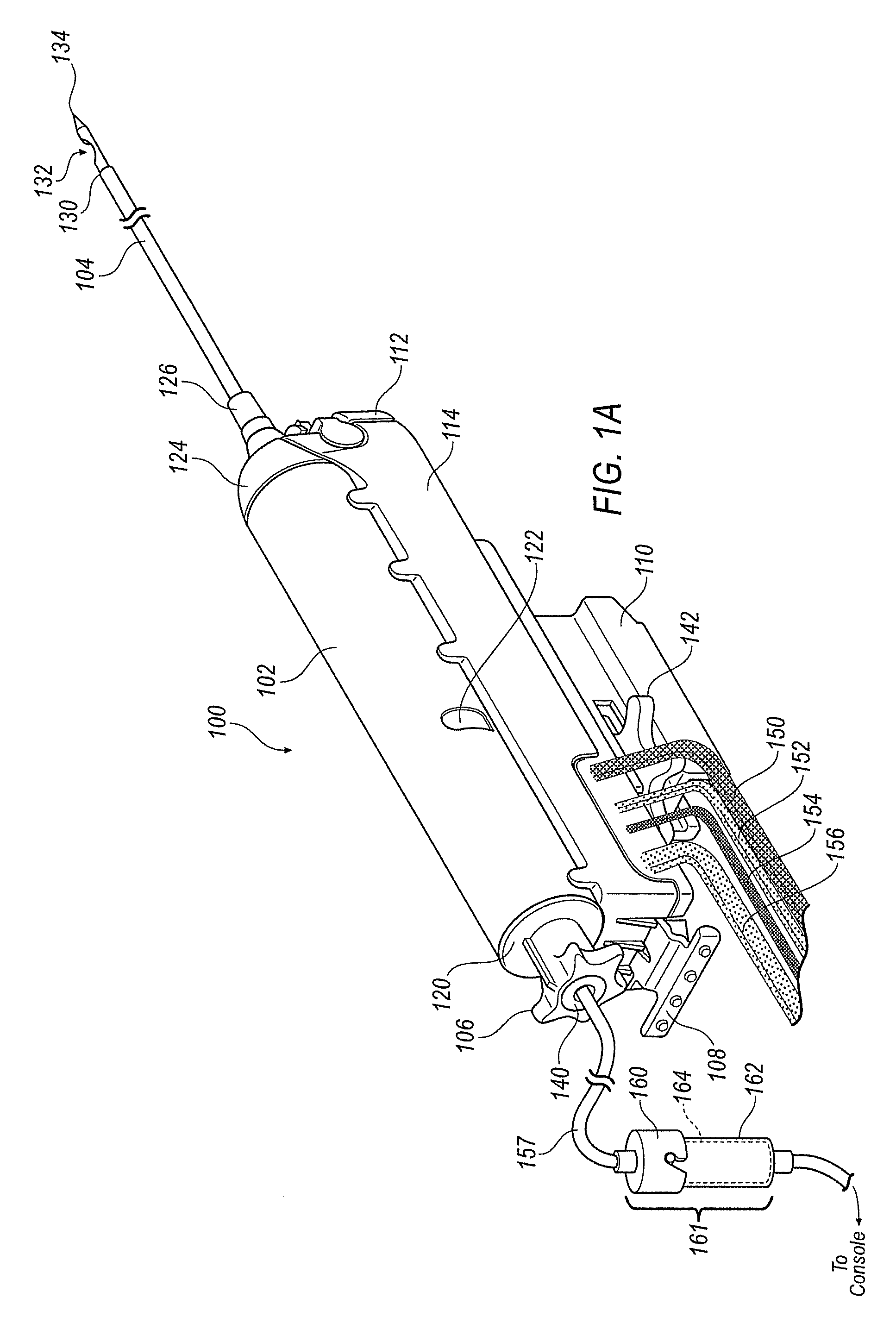
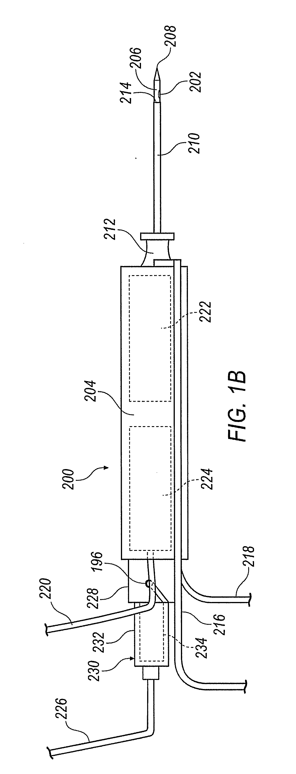
[0039]Referring now to the drawings, FIG. 1A illustrates a surgical device 100. Surgical device 100 is configured as a stereotactic type surgical device such as shown in commonly owned U.S. patent application Ser. No. 11 / 132,034. A handheld surgical device 200, such as shown in commonly owned U.S. patent application Ser. No. 10 / 970,269, the contents of which are incorporated herein by reference in its entirety, is shown in FIG. 1B and wil...


PUM


Abstract

A surgical device is disclosed that comprises a cutting element, a biopsy line, a motor line, and a vacuum line. The cutting element includes an outer cannula and an inner cannula. The biopsy line is operatively connected to a control console for moving the inner cannula within the outer cannula to cut tissue cores. The motor line is operatively connected to the control console for operating the surgical device. The vacuum line is operatively connected to the inner cannula. A selectively openable tissue filter is operatively connected to the inner cannula, and is also connected to the vacuum line.

Description

BACKGROUND[0001]This disclosure relates to biopsy systems and methods for talking a biopsy. More specifically, this disclosure relates to biopsy systems for removing multiple tissue samples.[0002]In the diagnosis and treatment of breast cancer, it is often necessary to remove multiple tissue samples from a suspicious mass. The suspicious mass is typically discovered during a preliminary examination involving visual examination, palpitation, X-ray, MRI, ultrasound imaging or other detection means. When this preliminary examination reveals a suspicious mass, the mass must be evaluated by taking a biopsy in order to determine whether the mass is malignant or benign. Early diagnosis of breast cancer, as well as other forms of cancer, can prevent the spread of cancerous cells to other parts of the body and ultimately prevent fatal results.[0003]A biopsy of the breast, for example, can be performed by either an open procedure or a percutaneous method. The open surgical biopsy procedure fi...

Claims


Application Information

Patent Timeline
Patent Type & Authority Applications(United States)
IPC IPC(8): A61B10/02
CPCA61B10/0275A61B10/0283A61B2010/0093A61B2019/464A61B2010/0225A61B2017/00477A61B2010/0208A61B2090/064
Inventor HOFFA, MICHAELMILLER, MICHAEL E.MARK, JOSEPH L.FLAGLE, JACOBSMITH, JUSTINBUTCHER, JASON M.
Owner SUROS SURGICAL SYST
Why Eureka
Try Eureka