Biomarkers for detection of early- and late-stage endometrial cancer
a biomarker and endometrial cancer technology, applied in the field of cancer detection and treatment, can solve the problems of no reliable serum biomarker in clinical use, and no endometrial cancer has been reported
- Summary
- Abstract
- Description
- Claims
- Application Information
AI Technical Summary
Benefits of technology
Problems solved by technology
Method used
Image
Examples
example 1
Identification of Biomarkers for Ovarian Cancer using Strong Anion-Exchange ProteinChips
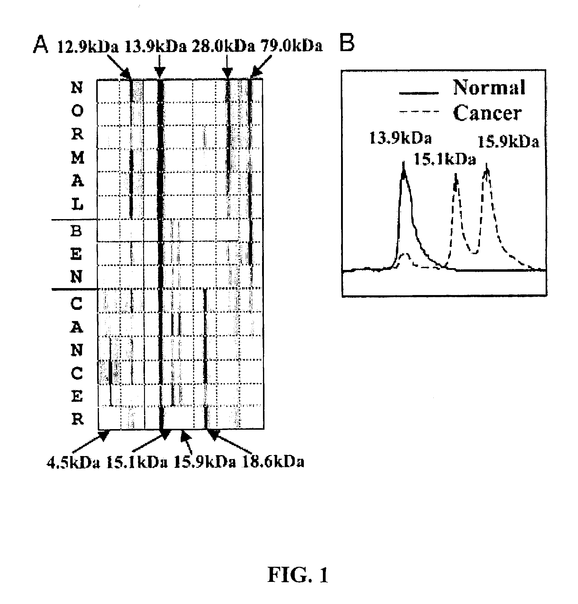
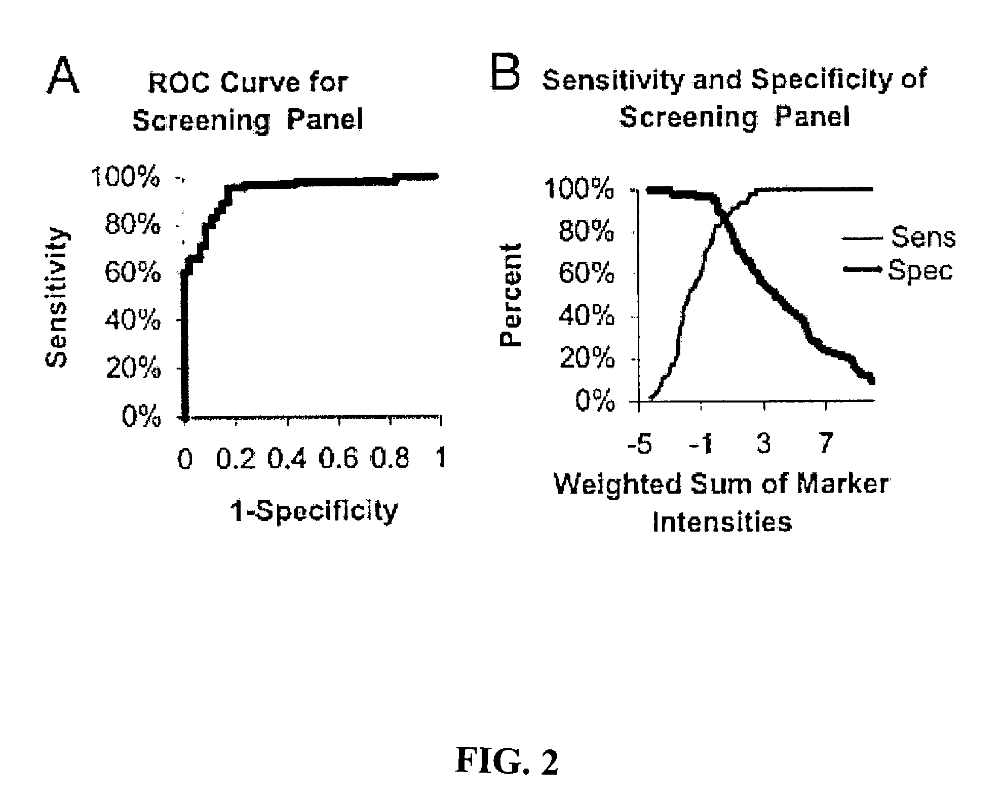
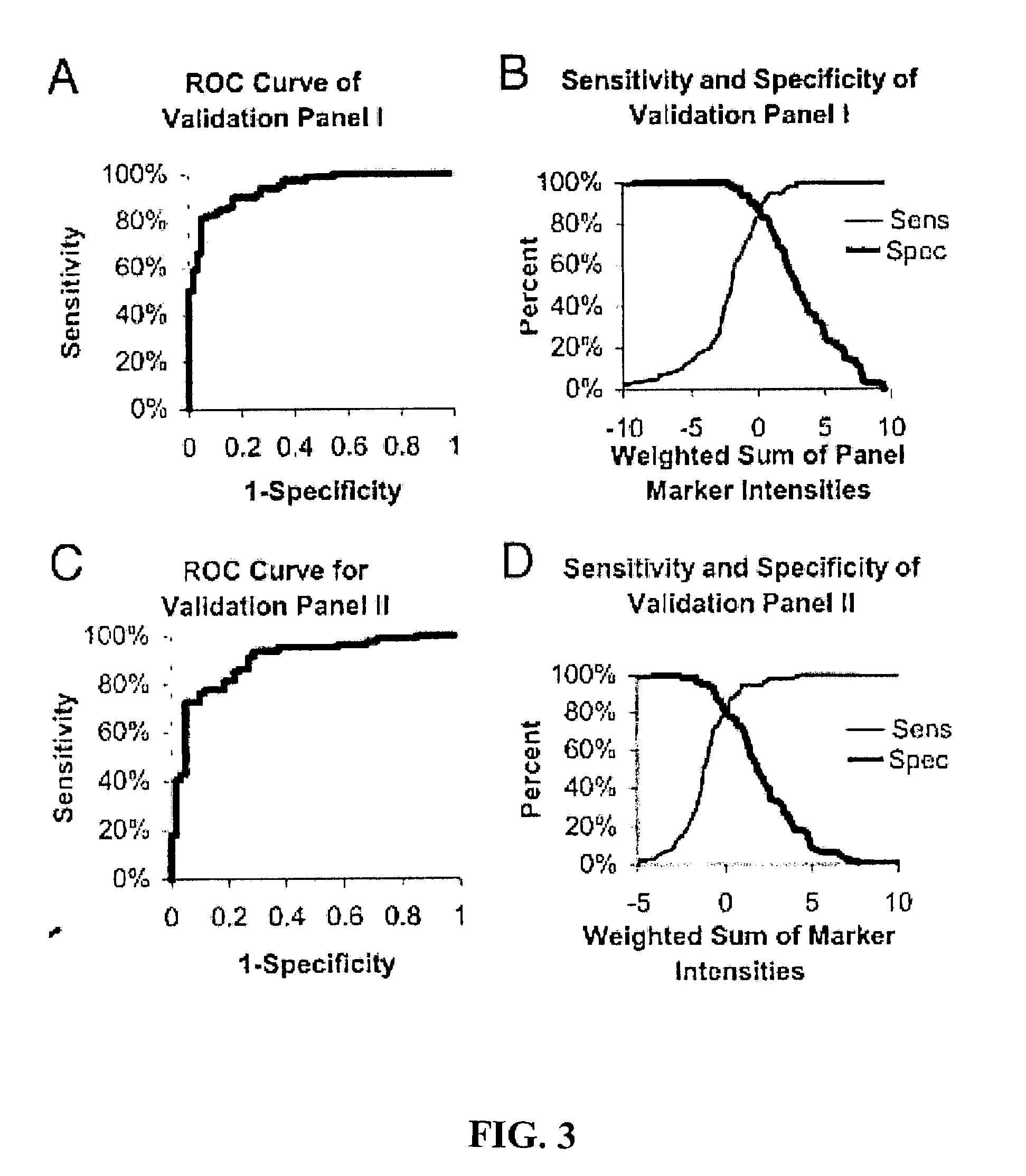
[0152]This example demonstrates three ovarian cancer biomarker protein panels that, when used together, effectively distinguished serum samples from healthy controls and patients with either benign or malignant ovarian neoplasia. In summary, 184 serum samples from patients with ovarian cancer (n=109), patients with benign tumors (n=19), and healthy donors =56) were analyzed on strong anion-exchange surfaces using surface-enhanced laser desorption / ionization time-of-flight mass spectrometry technology. Univariate and multivariate statistical analyses applied to protein-profiling data obtained from 140 training serum samples identified three biomarker protein panels. The first panel of five candidate protein biomarkers, termed the screening biomarker panel, effectively diagnosed benign and malignant ovarian neoplasia (95.7% sensitivity, 82.6% specificity, 89.2% accuracy, and receiver operating char...
example 2
Characterization of Ovarian Cancer Serum Biomarkers for Early Detection
[0164]This example demonstrates the identification, characterization, and validation of the proteins that represent the SELDI-TOF-MS peaks from the ovarian cancer biomarker panels. Mass spectrometry and other analytical methods were employed to identify proteins of interest in human serum. The Example describes the identity of proteins that represent the m / z peaks 12.9, 13.8, 15.1, 15.9, 28 and 78.9 kDa in the ovarian cancer biomarker panels. Using micro-liquid chromatography-tandem mass spectrometry, the following m / z peaks were identified as: transthyretin (TTR): 12.9 kDa and 13.9 kDa, hemoglobin, both alpha-hemoglobin (alpha-Hb): 15.1 kDa, and beta-hemoglobin (beta-Hb): 15.9 kDa, apolipoprotein AI (ApoAI): 28 kDa and transferrin (TF): 78.9 kDa. Western and ELISA techniques (independent of SELDI) confirmed the differential expression of TTR, Hb and TF in a group of ovarian cancer serum samples. Multivariate ana...
example 3
Identification of Additional Ovarian Cancer Serum Biomarkers
[0202]This example demonstrates the identification of additional biomarkers from serum samples that exhibit sensitivity and specificity in detecting ovarian neoplasia. These biomarkers were identified using SELDI as described above. The following table lists the protein identity, m / z ratios (in Daltons, “Marker”), cut point, sensitivity (“Sens”), specificity (“Spec”), accuracy (“Acc”) of each biomarker. Subsequent columns indicate the mean level observed for each biomarker in the screening (normal and neoplasm) and validation (nonmalignant and malignant) panels. N=140.
TABLE 5Mean Level of BiomarkerValidationCutScreeningNon-ProteinMarkerPointSensSpecAccNormalNeoplasmMalignantMalignantM19531.180.700.630.671.574.252.953.67M20651.700.630.700.661.733.232.632.81M22160.870.650.630.641.042.601.812.30M29281.350.550.830.690.792.061.381.84M29371.790.690.720.701.813.352.143.36M31431.590.650.650.651.642.592.022.47M34230.480.570.740.660....
PUM
 Abstract
Description
Claims
Application Information
 - R&D
- Intellectual Property
- Life Sciences
- Materials
- Tech Scout
- Unparalleled Data Quality
- Higher Quality Content
- 60% Fewer Hallucinations
Browse by: Latest US Patents, China's latest patents, Technical Efficacy Thesaurus, Application Domain, Technology Topic, Popular Technical Reports.
© 2025 PatSnap. All rights reserved.Legal|Privacy policy|Modern Slavery Act Transparency Statement|Sitemap|About US| Contact US: help@patsnap.com


