
Method and Apparatus for Quantitative Analysis of Breast Density Morphology Based on MRI

a breast density and quantitative analysis technology, applied in image analysis, image enhancement, instruments, etc., can solve the problems of higher cancer incidence, affecting cancer risk, and showing association, and achieve the effect of facilitating the investigation of the relationship

Active Publication Date: 2011-06-23
RGT UNIV OF CALIFORNIA
View PDF10 Cites 21 Cited by
  • Summary
  • Abstract
  • Description
  • Claims
  • Application Information

AI Technical Summary

Benefits of technology

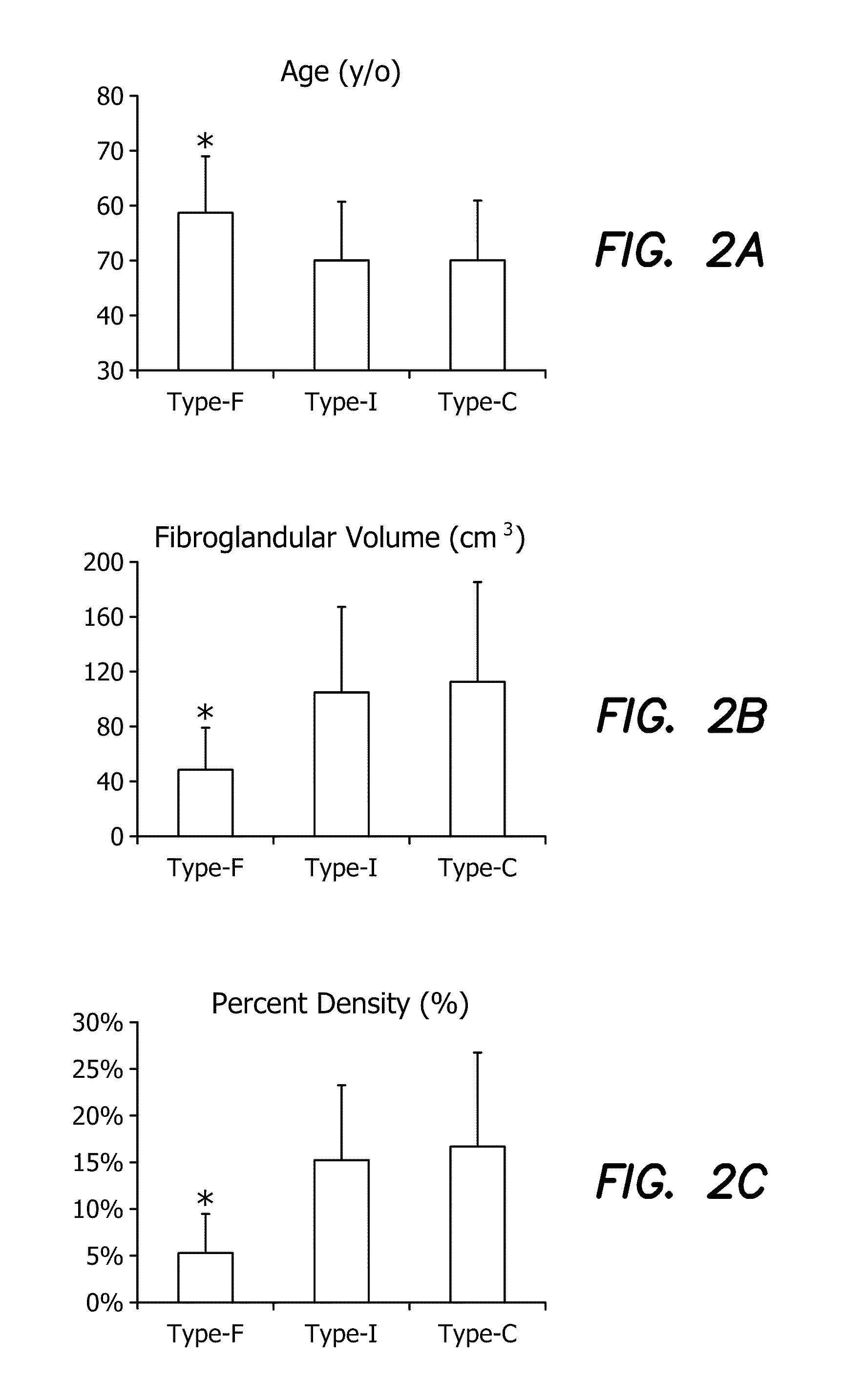
[0015]The results suggest that these morphological parameters analyzed from three dimensional MRI can be used to distinguish between intermingled and central dense tissue distribution patterns, and hence may be used to characterize breast parenchymal pattern quantitatively. The availability of these quantitative morphological parameters may facilitate the investigation of the relationship between parenchymal pattern and breast cancer risk.

Problems solved by technology

Increased density over time has also been shown to be associated with higher cancer incidence.
The adipose tissue that is abundantly present around the ductal epithelium of the mammary gland may function as a slow release depot for lipid-soluble carcinogenic agents, and thus may affect cancer risk.
However, the relationship between parenchymal pattern and cancer risk has never been reported, possibly due to the lack of both the imaging modality necessary to reveal the distribution pattern and the appropriate analysis methods.
However, as these features arise in part from the way that tissues overlap on the projected image, the analyzed parameters may be affected by different positioning of the breast and the degrees of compression.

Method used


Image

  • Method and Apparatus for Quantitative Analysis of Breast Density Morphology Based on MRI
  • Method and Apparatus for Quantitative Analysis of Breast Density Morphology Based on MRI
  • Method and Apparatus for Quantitative Analysis of Breast Density Morphology Based on MRI

Examples


Embodiment Construction

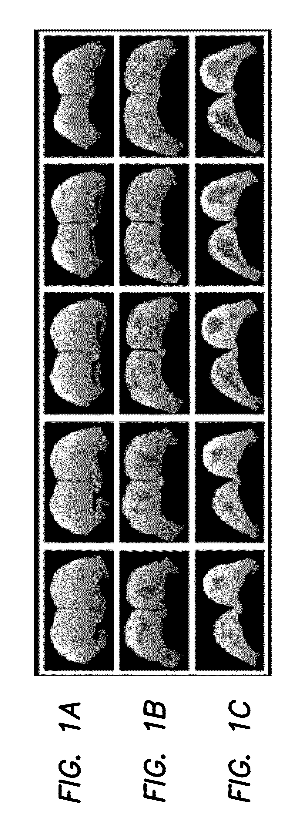
[0051]We have previously published an analysis method utilizing computer algorithms to segment the fibroglandular tissue for quantitative measurement of the percent density in the whole breast using MRI. In the illustrated embodiment we address a new question: In addition to the percent density, we use quantitative parameters to characterize the distribution pattern of the dense tissues. As an initial approach, we analyzed two distinct breast parenchymal patterns that can be classified visually: The intermingled pattern with intermixed fatty and fibroglandular tissues, and the central pattern with confined fibroglandular tissue inside surrounded by fatty tissue outside. Breasts from these two groups may have comparable percent densities, but differ in the distribution pattern of their dense tissue. Four different morphological parameters were calculated based on the three dimensional distribution pattern of segmented fibroglandular tissues, and their capacity to differentiate betwee...


PUM


Abstract

A method and apparatus configured to analyze breast density based on magnetic resonance imaging (MRI) of a breast of a patient includes the steps of segmenting an MR image of the breast from one set of three-dimensional breast MRI images, and analyzing the amount of dense tissue and the morphological distribution of the dense tissue and a data processor configured by software to perform these steps. Analyzing the amount of dense tissue and the morphological distribution of the dense tissue includes the steps of segmenting tissue data to separate breast tissue from other body tissue, separating tissue data of the dense and fatty tissues in the breast, and analyzing the morphological distribution of dense tissue in the breast to derive one or more three dimensional morphological parameters of the dense tissue distribution.

Description

RELATED APPLICATIONS[0001]The present application is related to U.S. Provisional Patent Application, Ser. No. 61 / 287,590, filed on Dec. 17, 2010, which is incorporated herein by reference and to which priority is claimed pursuant to 35 USC 119.BACKGROUND OF THE INVENTION[0002]1. Field of the Invention[0003]The invention relates to methodologies to analyze breast density based on magnetic resonance imaging (MRI).[0004]2. Description of the Prior Art[0005]Mammary gland architecture may play an important role in determining the susceptibility of developing breast cancer. The most well-studied parameter is mammographic density, evaluated as the percentage of dense tissue area over the total breast area on mammograms. There are numerous studies reporting mammographic density as a strong risk factor; the higher the percentage, the higher the breast cancer risk. Increased density over time has also been shown to be associated with higher cancer incidence. There is also evidence suggesting ...

Claims


Application Information

Patent Timeline
IPC IPC(8): G06K9/00
CPCG06T7/2026G06T2207/30068G06T2207/10088G06T7/0012G06T7/238
Inventor SU, MIN-YINGNIE, KE
Owner RGT UNIV OF CALIFORNIA
Learn More