system and method for real-time surface and volume mapping of anatomical structures

a real-time surface and volume mapping technology, applied in the field of system and method for real-time surface and volume mapping of anatomical structures, can solve the problems of increasing radiation exposure, prolonging the time of the procedure, and causing the patient to be prone to radiation exposur

Inactive Publication Date: 2011-06-30
ST JUDE MEDICAL INT HLDG SARL
View PDF17 Cites 91 Cited by
  • Summary
  • Abstract
  • Description
  • Claims
  • Application Information

AI Technical Summary

Problems solved by technology

One drawback of known cannulation methodologies is that the procedures such as this can be rather lengthy procedures that employ a “trial and error” method for searching for the ostium (i.e., the physician “pokes” or probes around the surface until the ostium is found).
Without information regarding where the physician previously poked or probed, the same area may be poked or probed several times, thereby lengthening the time of the procedure and increasing the amount of radiation exposure (i.e., x-ray) needed, thus resulting in exposing the anatomical structure or region of interest to redundant irritation.
Lengthy procedure times

Method used


Image

  •  system and method for real-time surface and volume mapping of anatomical structures
  •  system and method for real-time surface and volume mapping of anatomical structures
  •  system and method for real-time surface and volume mapping of anatomical structures

Examples


Embodiment Construction

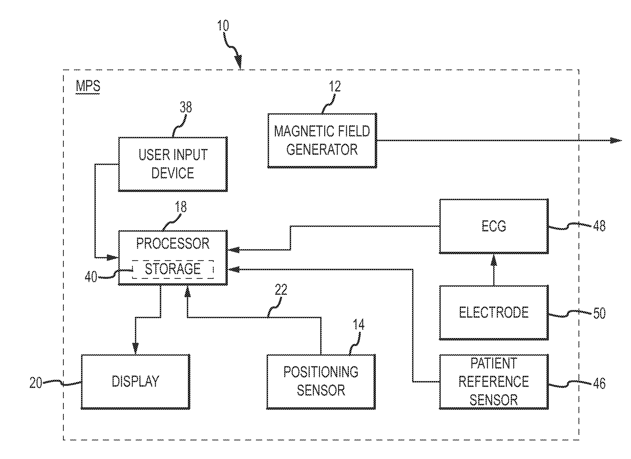
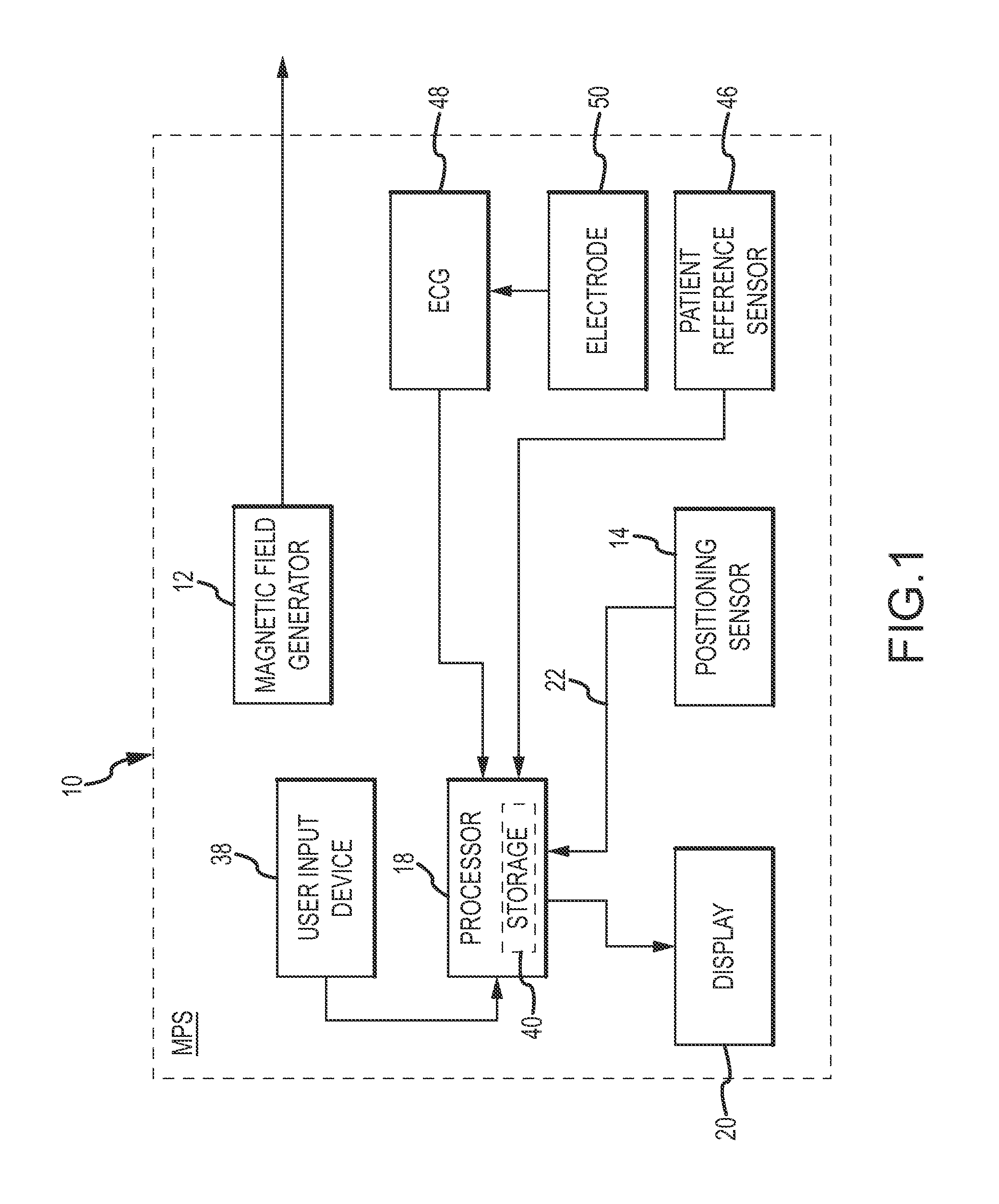
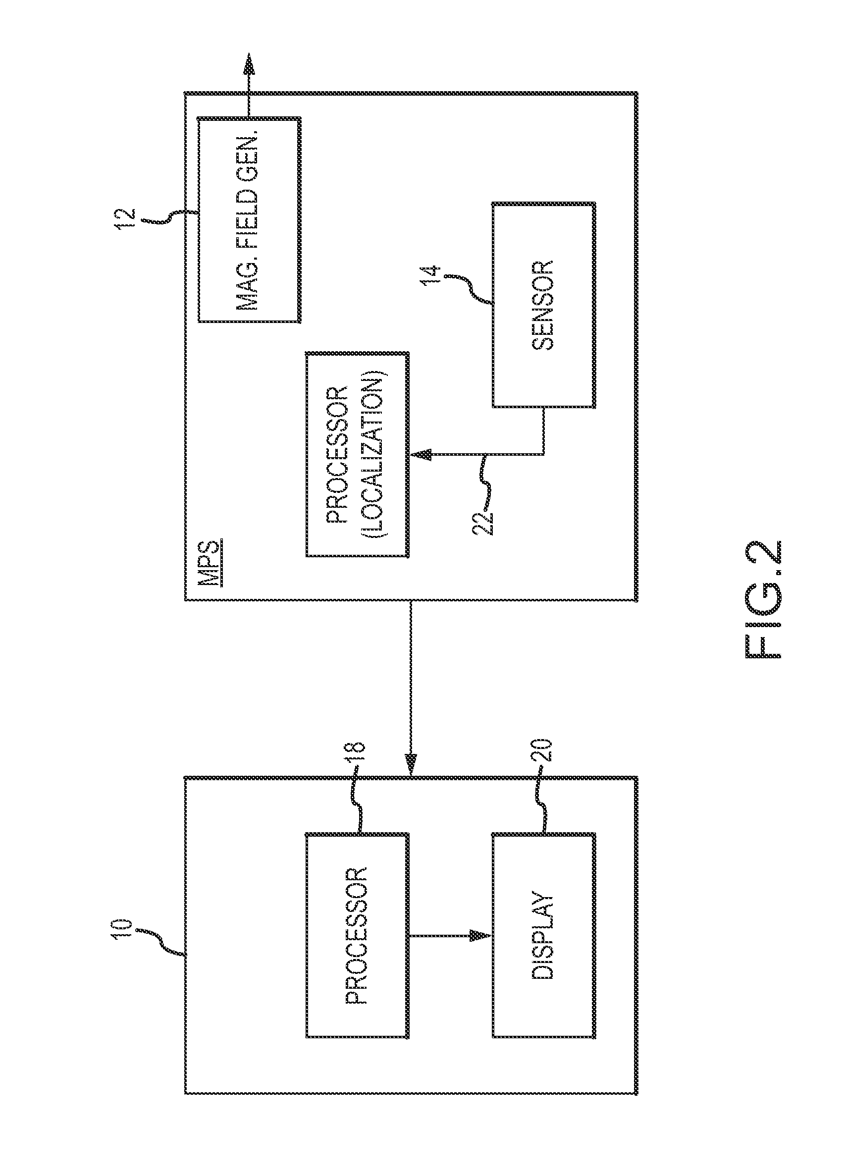
[0026]Referring now to the drawings wherein like reference numerals are used to identify identical components in the various views, FIG. 1 illustrates an exemplary embodiment of a system 10 for mapping a volume of an anatomical structure within a region of interest (ROI) of a patient's body. In an exemplary embodiment, the system 10 comprises a medical positioning system (MPS). It should be noted, however, that in other exemplary embodiments, such as that illustrated in FIG. 2, rather than comprising an MPS, the system 10 is separate and distinct from the MPS but configured for use in conjunction with an MPS.

[0027]In either embodiment, the MPS is configured to serve as a localization system, and therefore, is configured to determine positioning (localization) data with respect to one or more MPS sensors and to output a respective location reading. The location readings may each include at least one or both of a position and an orientation (P&O) relative to a reference coordinate sys...


PUM


Abstract

A method and system for mapping a volume of an anatomical structure includes a processor for computing a contour of a medical device as a function of positional and/or a shape constraints, and to translate the contour into known and virtual 3D positions. The processor is configured to determine a spatial volume based on a virtual position, and to render a 3D representation of the spatial volume. A method and system of mapping a surface of an anatomical structure includes a processor configured to obtain an image of the structure. The processor is further configured to receive a signal indicative of a medical device contacting the surface of the anatomical structure, and to determine a position of the device upon when contact has been made. The processor is configured to superimpose marks on the image indicative of a contact points between the device and the structure.

Description

BACKGROUND OF THE INVENTION[0001]a. Field of the Invention[0002]The present disclosure relates to a system and method for real-time surface and volume mapping of an anatomical structure.[0003]b. Background Art[0004]In a cannulation procedure, a physician cannulates into a vessel of an anatomical structure, such as, for example, a vessel of the heart. In such procedures, a physician or clinician inserts a medical device or tool, such as, for example, a catheter, into an insertion region of a patient, which, in an exemplary embodiment, may comprise the Superior Vena Cava (SVC) of the heart. Once the medical device or tool is inserted, the physician or clinician uses the device or tool to probe around a surface of the anatomical structure searching for the ostium of the vessel the physician is attempting to cannulate through.[0005]One drawback of known cannulation methodologies is that the procedures such as this can be rather lengthy procedures that employ a “trial and error” method f...

Claims


Application Information

Patent Timeline
IPC IPC(8): A61B6/00
CPCA61B5/06A61B5/064A61B5/7285A61B5/7289A61B6/541G06T2219/008G06T19/00G06T2207/10072G06T2207/30101G06T2210/41G06T7/2033A61B5/7425A61B5/743G06T7/246
Inventor COHEN, AMITKARIV, ITAY
Owner ST JUDE MEDICAL INT HLDG SARL
Why Eureka
Try Eureka