Self-administered breast ultrasonic imaging systems

a breast ultrasound and self-administering technology, applied in the field of medical devices and medical diagnostics, can solve the problems of inconvenient use, increased labor intensity, and increased labor intensity, and achieves the effects of saving unnecessary travel and strain on the patient, high utility, and high survival ra

Inactive Publication Date: 2011-12-08
ANITE DORIS NKIRUKA
View PDF5 Cites 38 Cited by
  • Summary
  • Abstract
  • Description
  • Claims
  • Application Information

AI Technical Summary

Benefits of technology

[0012]Alternatively, even in the absence of a network link to a remote skilled interpreter, an automated or semi-automated device and method used to produce skilled medical images could still be of high utility even when used in a mode where a patient might have a scan done in a remote location or after hours setting, and the medical image either printed out or saved in a computer memory storage device for subsequent analysis by a skilled interpreter. This way the

Problems solved by technology

Ultrasonic imaging equipment, although certainly not inexpensive, also contrasts favorably in expense to other much more expensive forms of safe medical imaging technology, such as magnetic resonance imaging (MRI) methods.
One problem with ultrasonic imaging technology, however is that this technology still requires that a skilled operator be present to move the ultrasonic transducer about the body, and interpret the images on the video monitor of the computerized ultrasonic imaging equipment.
This need for a skilled operator is unfortunate, because due to advances in electronics and instrumentation, the basic costs of medical ultrasonic imaging equipment itself is likely to continue to substantially decrease over the coming years.
However since skilled operators are scarce, these equ

Method used


Image

  • Self-administered breast ultrasonic imaging systems
  • Self-administered breast ultrasonic imaging systems
  • Self-administered breast ultrasonic imaging systems

Examples


Embodiment Construction

[0035]Here ultrasonic transducers will occasionally be referred to in the alternative as the ultrasonic probe, ultrasonic probe head, or occasionally the probe head or probe.

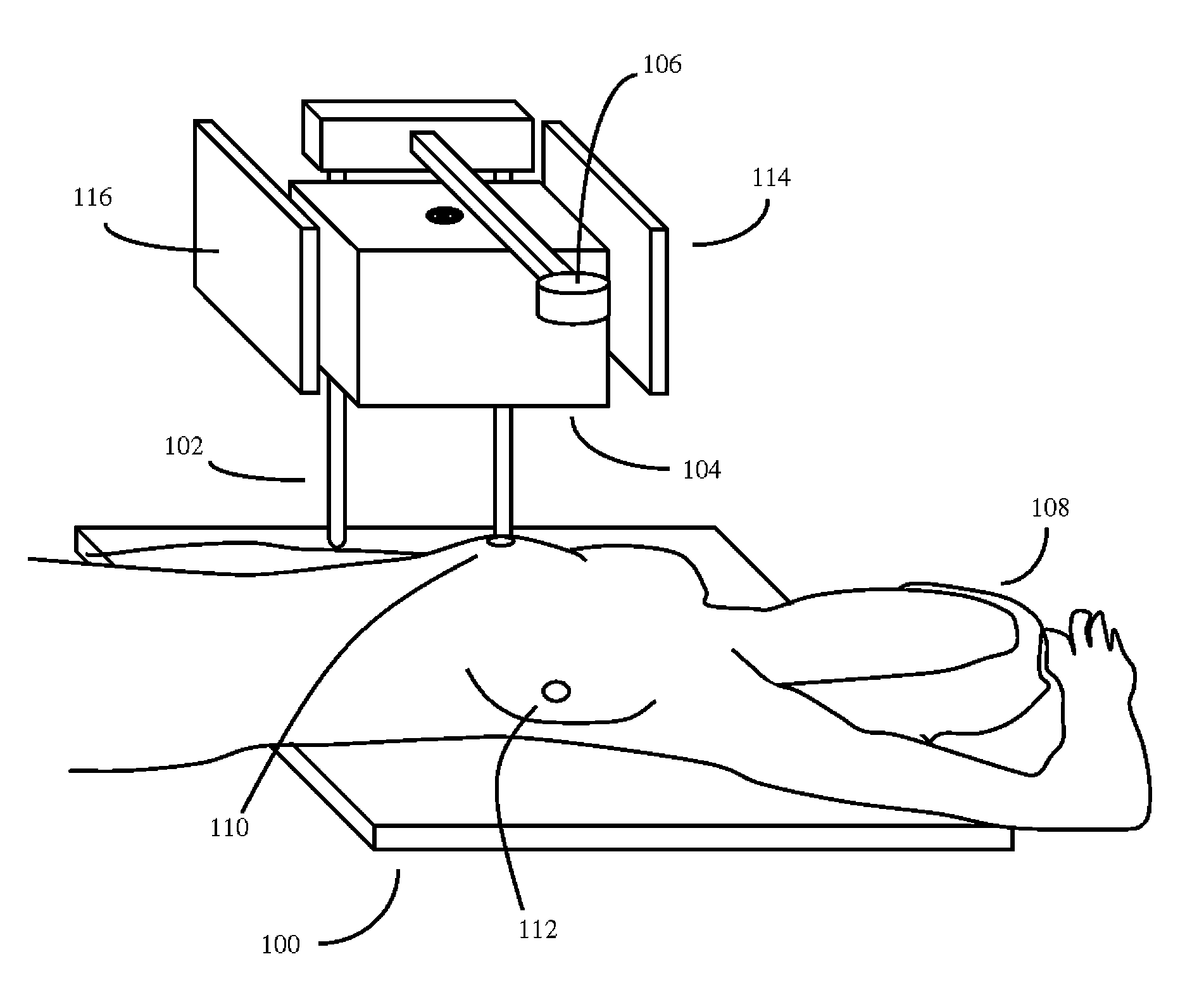
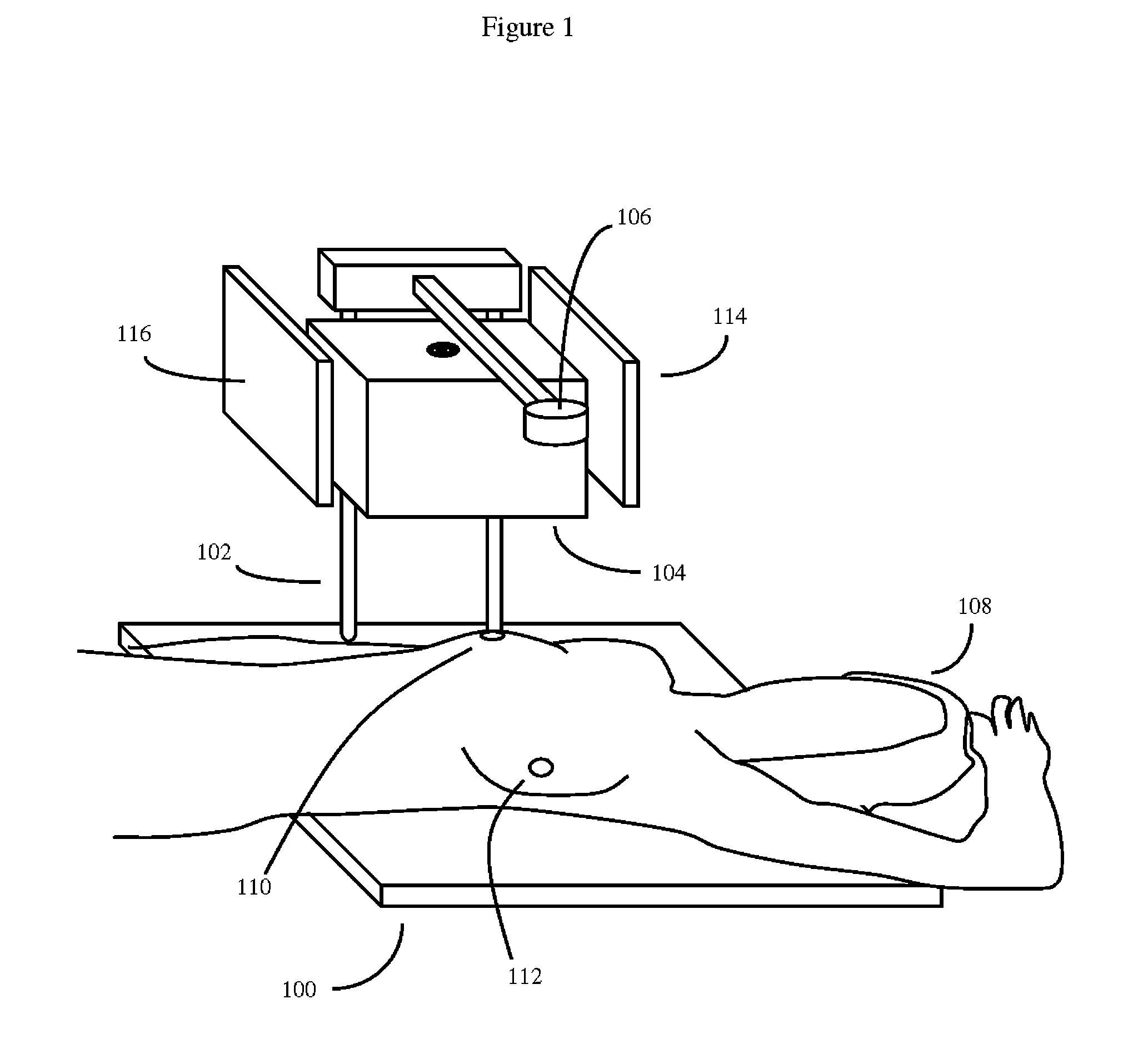
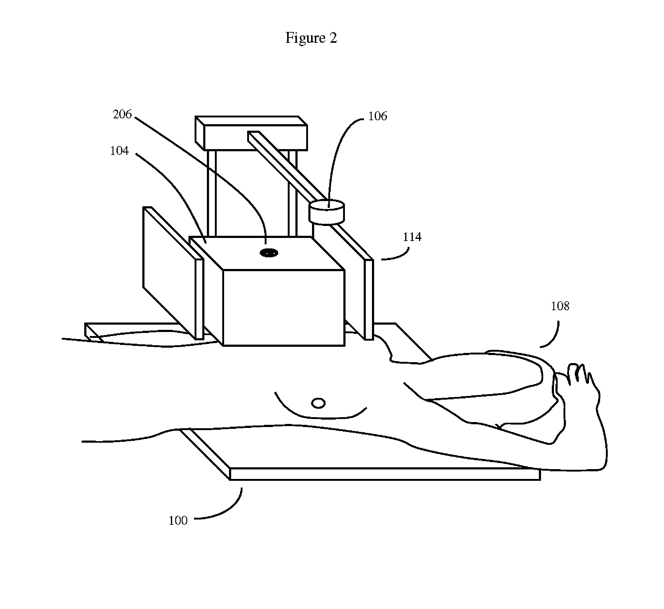
[0036]As previously discussed, the invention may be viewed as a medical ultrasonic transducer guiding system capable of being used by unskilled users. This system will generally comprise at least a moveable ultrasonic transducer guidance platform configured to enable an ultrasonic transducer to be repositioned in multiple positions about a patient's breast (either one breast at a time, or both breasts at a time), and produce useful medical diagnostic images of the patient's breast.

[0037]This guidance platform may optionally also have or comprise sensors that detect the position of the patient, the patient's breast, and the ultrasonic transducer. These sensors may include image sensors, as well as ultrasonic transducer position sensors that may be built into the actuators that move the ultrasonic transducer, or m...


PUM


Abstract

An ultrasonic breast examination system suitable for use by unskilled users, comprising a patient positioning platform with a patient positioning image sensor, and a moveable ultrasonic transducer guidance device. The transducer guidance device can be a fluid filled container with a flexible side or bottom, and a mechanism to automatically move an ultrasonic transducer over the breast. The system will be controlled by at least one microprocessor, associated software, and an optional touch sensitive display screen. The system may use an image sensor to properly position the patient so that the transducer guidance device may be properly positioned proximate to the patient's breast. The touch sensitive display screen is designed to allow the system to be directly operated by an unskilled patient. The system will often be connected to a network, such as the Internet, so that remote operators may interpret breast ultrasound images and optionally control the system.

Description

CROSS REFERENCE TO RELATED APPLICATIONS[0001]This application is a continuation in part of U.S. patent application Ser. No. 12 / 826,188, “Self-administered Medical Ultrasonic Imaging systems, filed on Jun. 29, 2010, Dr. Doris Nkiruka Anite inventor; U.S. application Ser. No. 12 / 826,188 in turn claims the priority benefit of Nigerian patent application NG / P / 2010 / 374, entitled “SELF-ADMINISTERED DIAGNOSTIC MEDICAL DEVICES AND SOFTWARE”, filed on Jun. 4, 2010, and U.S. Provisional Application No. 61 / 357,953, filed on Jun. 23, 2010, also entitled “SELF-ADMINISTERED DIAGNOSTIC MEDICAL DEVICES AND SOFTWARE”, both by Dr. Doris Nkiruka Anite inventor.BACKGROUND OF THE INVENTION[0002]1. Field of the Invention[0003]The application is in the general field of medical devices and medical diagnostics, particularly ultrasonic imaging technology.[0004]2. Description of the Related Art[0005]Medical imaging technology, in particular ultrasonic medical imaging technology, has revolutionized the diagnos...

Claims


Application Information

Patent Timeline
IPC IPC(8): A61B8/14
CPCA61B8/0825A61B8/4209A61B8/4263A61B8/4281A61B8/565A61B8/4461A61B8/4472A61B8/461A61B8/467A61B8/429A61B8/465
Inventor ANITE, DORIS NKIRUKA
Owner ANITE DORIS NKIRUKA
Why Eureka
Try Eureka