Method and Apparatus for Segmenting Biological Cells in a Picture

Inactive Publication Date: 2011-12-29
FRAUNHOFER GESELLSCHAFT ZUR FOERDERUNG DER ANGEWANDTEN FORSCHUNG EV
View PDF2 Cites 6 Cited by
  • Summary
  • Abstract
  • Description
  • Claims
  • Application Information

AI Technical Summary

Benefits of technology

[0029]In accordance with further embodiments of the present invention, the second fast marching algorithm may be configured such that a meeting line between a first fast marching front, which starts from a first cell center of a first biological cell, and a second fast marching front, which starts from a second cell center of a second biological cell, forms a boundary between the first biological cell and the second biological cell. By forming boundaries between different cells on the basis of meeting lines of fast marching fronts of the second fast marching algorithm, over-segmentation of cells, as it may be the case, for example, in the above-mentioned watershed algorithm, c

Problems solved by technology

In particular in bone marrow smears, cells (typically white blood corpuscles—leukocytes) are mostly exist in the form of cell clusters (cell groups), i.e. the individual cells are directly adjacent to one another and are therefore diffi

Method used


Image

  • Method and Apparatus for Segmenting Biological Cells in a Picture
  • Method and Apparatus for Segmenting Biological Cells in a Picture
  • Method and Apparatus for Segmenting Biological Cells in a Picture

Examples


Example

[0046]Before embodiments of the present invention will be explained in more detail below with reference to the accompanying figures, it shall be noted that elements that are identical or identical in function will be designated by the same reference numerals, and that repeated description of said elements will be dispensed with. Therefore, descriptions of elements having identical reference numerals are interchangeable.

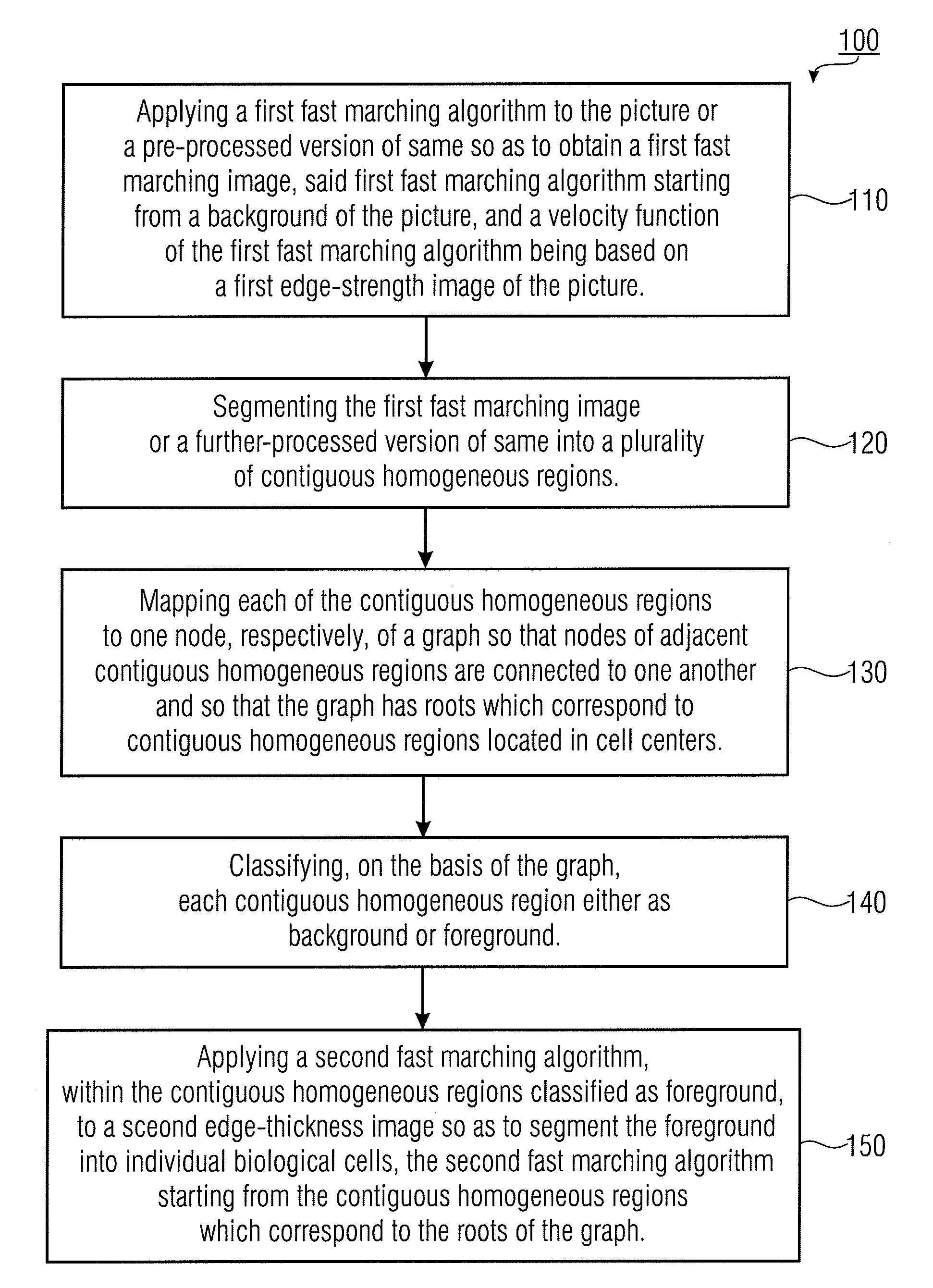
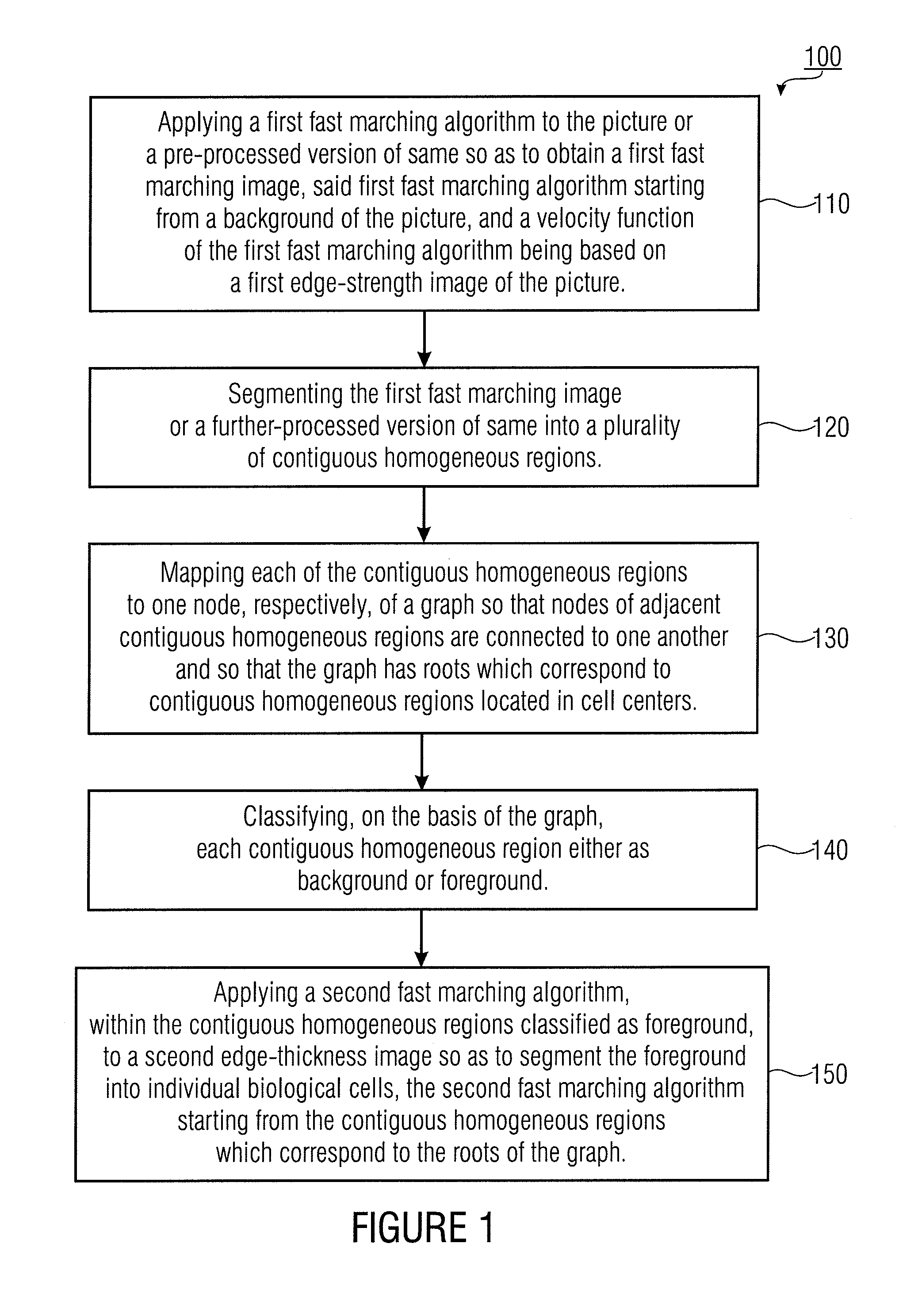
[0047]FIG. 1 shows a flowchart of a method 100 of segmenting biological cells in a picture, so that the biological cells represent a foreground of the picture, in accordance with an embodiment of the present invention. A biological cell may be a white blood corpuscle (leukocyte), a red blood corpuscle (erythrocyte) or any other biological cell. For example, the picture may comprise a plurality of biological cells of different cell types. The cells may be present in so-called cell clusters (cell groups), for example. The picture may be an RGB (red / green / blue) image, a ...


PUM


Abstract

A method of segmenting biological cells in a picture so that the biological cells represent a foreground of the picture includes a step of applying a first fast marching algorithm to the picture or to a pre-processed version of same in order to obtain a first fast marching image. In addition, the method includes a step of segmenting the first fast marching image or a further-processed version of same into a plurality of homogeneous regions. Furthermore, the method includes a step of mapping each of the homogeneous regions to one node of a graph, respectively. In addition, the method includes a step of classifying each homogeneous region either as background or foreground on the basis of the graph. Moreover, the method includes a step of applying a second fast marching algorithm within the homogeneous regions classified as foreground so as to segment the foreground into individual biological cells.

Description

CROSS-REFERENCE TO RELATED APPLICATIONS[0001]This application claims priority to German Patent Application No. 102010024859.2 filed 24 Jun. 2010, which is incorporated herein by reference.[0002]Embodiments of the present invention relate to a method and an apparatus for segmenting biological cells in a picture, as may be used, for example, for detecting and segmenting leukocytes in blood smears or bone marrow smears.BACKGROUND OF THE INVENTION[0003]One important component of hematology is the differential blood count. Systems from the field of “computer-assisted microscopy” (CAM) enable automatic analysis of blood smears and support hematologists in classifying cells, and thus they form a supplement for modern hematological laboratory diagnostics. It is in a fast, precise and highly efficient manner that modern hematology systems provide important information about the cell population of peripheral blood. However, up to 40% of samples may subsequently be manually differentiated unde...

Claims


Application Information

Patent Timeline
IPC IPC(8): G06K9/34
CPCG06T7/0081G06T7/0093G06T2207/20161G06T2207/30024G06T2207/20144G06T2207/10056G06T7/11G06T7/162G06T7/194G06V20/695
Inventor HASSLMEYER, ERIKELTER, MATTHIASZERFASS, THORSTENSCHLARB, TIMO
Owner FRAUNHOFER GESELLSCHAFT ZUR FOERDERUNG DER ANGEWANDTEN FORSCHUNG EV
Why Eureka
Try Eureka