Determination of a measure of a glycation end-product or disease state using tissue fluorescence in combination with one or more other tests

a tissue fluorescence and end-product technology, applied in the field of disease state determination, to achieve the effect of high sensitivity, high sensitivity and high sensitivity

a tissue fluorescence and end-product technology, applied in the field of disease state determination, to achieve the effect of high sensitivity, high sensitivity and high sensitivity

US20120078075A1Inactive Publication Date: 2012-03-29MAYNARD JOHN D +1

Image

  • Determination of a measure of a glycation end-product or disease state using tissue fluorescence in combination with one or more other tests
  • Determination of a measure of a glycation end-product or disease state using tissue fluorescence in combination with one or more other tests
  • Determination of a measure of a glycation end-product or disease state using tissue fluorescence in combination with one or more other tests

Examples


example i

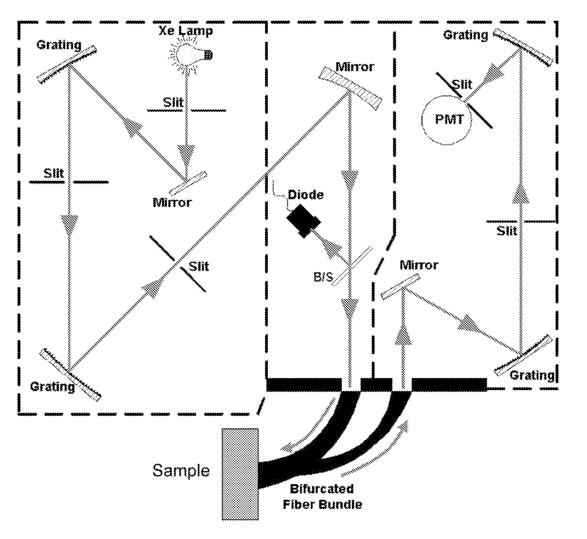
[0120 of such a system embodies a high-intensity arc lamp, shutter, monochromator and collimator as the core elements of the light source. The optical-coupling sub-system is comprised of a bifurcated fiber bundle that couples the excitation light to the tissue and collects fluorescence emanating from the tissue. The second leg of the bifurcated bundle couples the collected fluorescent light to the detection sub-system. The detection system contains a monochromator (separate from the monochromator of component A) and a detector such as a photomultiplier. The electrical signal corresponding to the tissue fluorescence is digitized, processed and stored by a computer (Component D). The computer also controls functions of other sub-systems such as the tuning of monochromators and opening closing shutters.

[0121]In Example II, the bifurcated fiber-optic bundle of Example I is replaced by a system of lenses and mirrors to convey excitation light from the light source to the tissue and then ...


PUM


Abstract

The present invention provides a method of determining disease state in an individual. A portion of the tissue of the individual is illuminated with excitation light, then light emitted by the tissue due to fluorescence of a chemical in the tissue responsive to the excitation light is detected. The HbA1c or FPG measurement of the individual (or other secondary indication of disease state) can also be determined. A model combining the tissue fluorescence and one or more of the secondary indications can be used to determine the disease state of the individual. In some embodiments, the tissue fluorescence can be used as an initial screen, and the combination with secondary indications only made for those individuals for whom the fluorescence screen indicates an increased likelihood of disease.

Description

CROSS REFERENCES TO RELATED APPLICATIONS[0001]This application claims priority to U.S. provisional application 61 / 420,444 filed Dec. 7, 2010; and claims priority as a continuation in part of U.S. application Ser. No. 11 / 964,675 filed Dec. 26, 2007; and as a continuation in part of U.S. application Ser. No. 11 / 561,380 filed Nov. 17, 2006, which application was a continuation of U.S. application Ser. No. 10 / 972,173 filed Oct. 22, 2004, now U.S. Pat. No. 7,139,598 issued Nov. 21, 2006, “Determination of a Measure of a Glycation End-Product or Disease State Using Tissue Fluorescence;” which was a continuation in part of U.S. application Ser. No. 10 / 116,272 filed Apr. 4, 2002, now U.S. Pat. No. 7,043,288 issued May 9, 2006, “Apparatus And Method For Spectroscopic Analysis Of Tissue To Detect Diabetes In An Individual,” and claimed the benefit of U.S. provisional application 60 / 515,343 filed Oct. 28, 2003, “Determination of a Measure of a Glycation End-Product or Disease State Using Tissu...

Claims


Application Information

Patent Timeline
29 Mar 2012
Publication
US20120078075A1
IPC
A61B5/145; A61B6/00
CPC
A61B5/0071; A61B5/0075; A61B5/443; A61B5/14546; A61B5/14532
Inventors
MAYNARD, JOHN D.; HULL, EDWARD