Magnetic field unit of an MRI system for image capturing a head region

a magnetic resonance tomography and magnetic field technology, applied in the field of magnetic field units of mri systems for image capture of head regions, can solve the problems of limited use of magnetic resonance tomography in the field of dentistry, high installation and operation costs, and limited dental image information of clinical relevance, so as to increase improve the quality of measuring signals

Active Publication Date: 2012-06-14
SIRONA DENTAL SYSTEMS
View PDF17 Cites 6 Cited by
  • Summary
  • Abstract
  • Description
  • Claims
  • Application Information

AI Technical Summary

Benefits of technology

[0035]Thus the coil for receiving radio frequencies is located as close as possible to the object to be scanned, namely the jaw region of a patient, which increases the quality of the measuring signals.
[0036]Advantageously, the magnetic field unit comprises a movable shielding device that can surround the magnetic field unit and the patient positioned therein such that only a minimum of radio frequencies from the environment can enter the region inside the shielding device. This increases the quality of the measuring signals.
[0037]A

Problems solved by technology

Very limited use has hitherto been made of magnetic resonance tomography in the field of dentistry.
This is primarily

Method used


Image

  • Magnetic field unit of an MRI system for image capturing a head region
  • Magnetic field unit of an MRI system for image capturing a head region
  • Magnetic field unit of an MRI system for image capturing a head region

Examples


Embodiment Construction

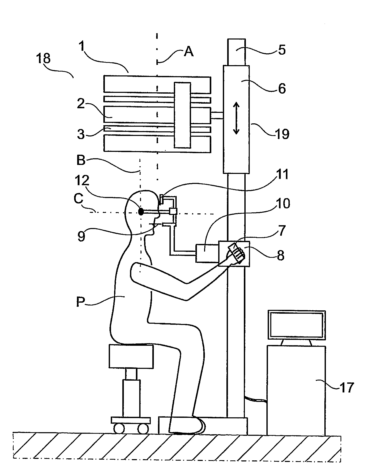
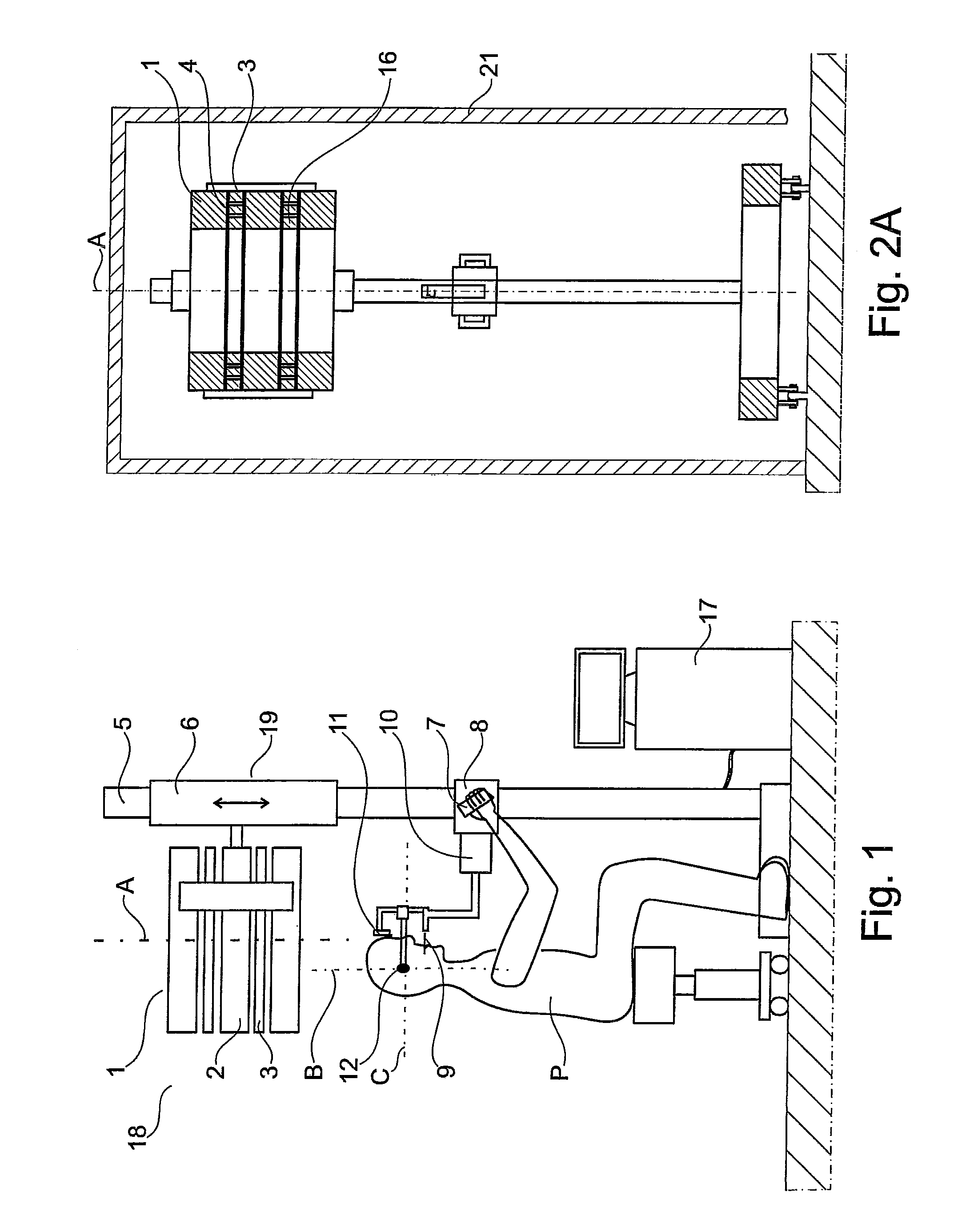
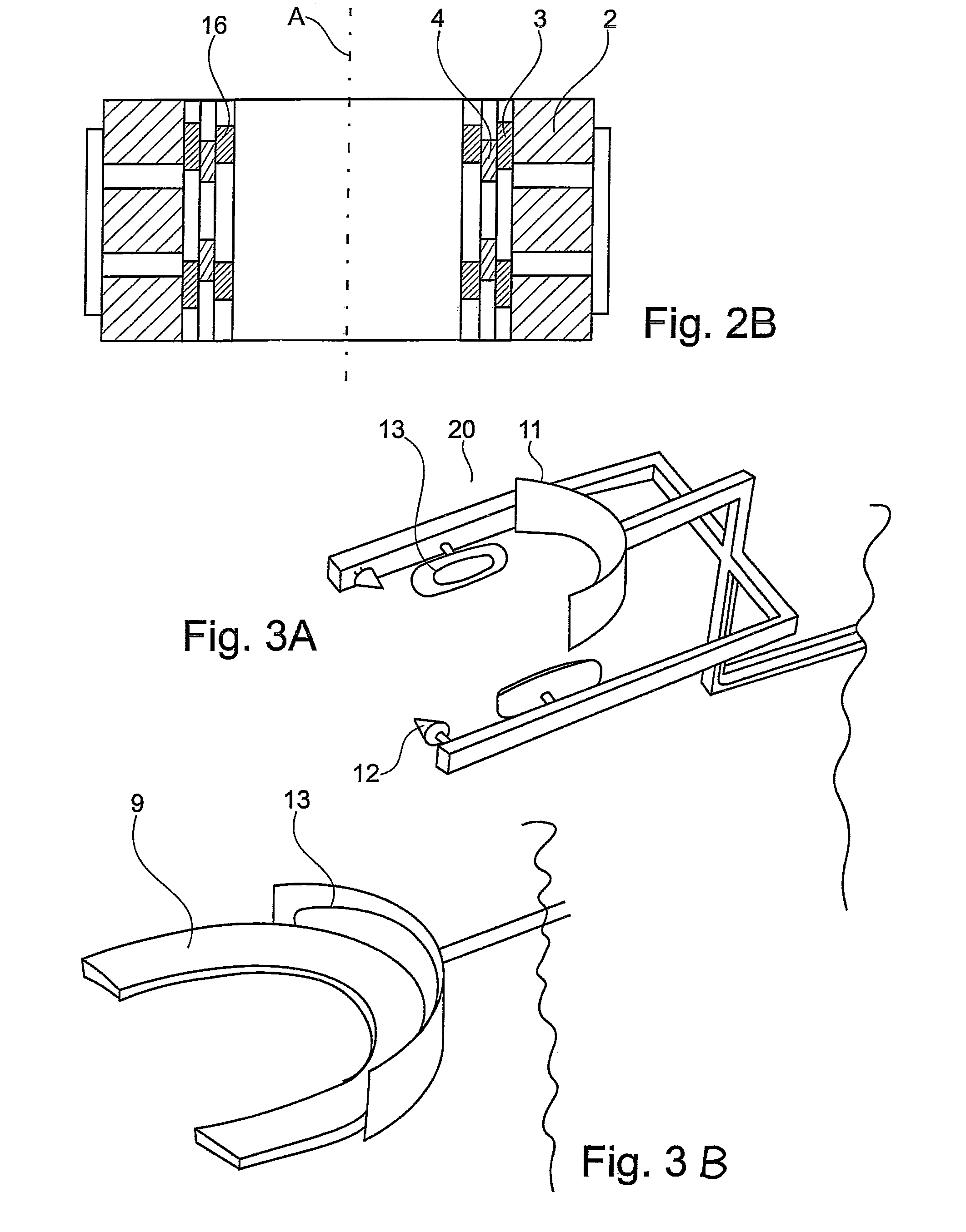
[0047]FIG. 1 is a side view of an MRT system 18 comprising a magnetic field unit 1 of the invention and a control unit 17. The magnetic field unit 1 comprises a plurality of permanent magnets 2 having a closed shape for the production of a main magnetic field and a device 3 consisting of a plurality of coils for the production of a gradient field, and coils 4 for generating and receiving radio frequencies. An example of an arrangement of the device 3 for the production of generating a gradient field and the coil 4 for generating and receiving radio frequencies is shown in FIG. 2A, which is a cross-sectional view of the magnetic field unit 1 of the invention. The coils of the device 3 for the production of a gradient field and the at least one coil 4 for generating and receiving radio frequencies are disposed in the space between the plurality of permanent magnets 2. The device 3 for the production of a gradient field and the at least one coil 4 for generating and receiving radio fre...


PUM


Abstract

The invention relates to a magnetic field unit (1) of an MRI system (18) for image capturing a head region as the region to be recorded, comprising at least one permanent magnet (2) for generating a main magnetic field, a device (3) for generating a gradient field, and at least one coil (4) for generating and receiving radio frequencies. The magnetic field unit (1) is fastened to a mounting (19) such that a longitudinal axis (A) of the magnetic field unit (1), together with a vertically extending axis, forms an angle of no more than +45° and that the magnetic field unit (1) and the patient (P) can be adjusted relative to each other such that the magnetic field unit (1) can be moved to a position surrounding the head of the patient (P).

Description

[0001]The present invention relates to a magnetic field unit of an MRT system for acquiring an image of a part of a head forming a region to be scanned, which magnetic field unit comprises at least one permanent magnet, a device for the production of a gradient field, and at least one coil for generating and receiving radio frequencies.PRIOR ART[0002]Very limited use has hitherto been made of magnetic resonance tomography in the field of dentistry. This is primarily due to the high cost of installation and operation, and the limited dental image information of clinical relevance.[0003]By contrast, X-ray images as can be produced by the device disclosed, for example, in EP 0 632 994 A1 are widely used. In this case, an X-ray diagnostic device for producing X-ray images of body parts, for example a jaw, is disclosed, in which a line detector camera and a radiation source disposed diametrically opposite thereto can be moved synchronously around the object to be scanned.[0004]The advant...

Claims


Application Information

Patent Timeline
IPC IPC(8): G01R33/44
CPCG01R33/3806G01R33/421G01R33/3875G01R33/383
Inventor RASCHE, VOLKERHELL, ERICHULRICI, JOHANNES
Owner SIRONA DENTAL SYSTEMS
Why Eureka
Try Eureka