Systems and methods for guiding catheters using registered images

a technology of registered images and catheters, applied in the field of system and method for guiding catheters, can solve the problems of inability to determine and register the true shape and configuration, the dynamic movement of a volume, and the current techniques, etc., to achieve the effect of guiding the physician to a three-dimensional view of the volume, providing two-dimensional information about the volume, and allowing the physician to see the volume in three-dimensional form

Inactive Publication Date: 2012-11-01
BOSTON SCI SCIMED INC
View PDF10 Cites 57 Cited by
  • Summary
  • Abstract
  • Description
  • Claims
  • Application Information

AI Technical Summary

Problems solved by technology

Current techniques, however, are incapable of determining and registering the true shape and configuration, as well as the dynamic movement of a volume, or at the least at a resolution high enough to provide a physician a comfortable understanding of the volume.
These devices only provide two-dimensional information about the volume, however, rather than the more preferred three-dimensional information.
In addition, not only do these device not give the physician a three-dimensional view of the volume, but also do not give an understanding of possible obstacles or movements within the volume itself, such as the opening and closing of valves, atrio-septal defects, atrio-septal defect closure plugs, and the like.
Some technologies are capable of generating and registered three-dimensional images, but these devices are typically incapable of producing a high resolution image of the interior space of the volume, since they operate from outside of the body, or from a location outside of the target volume itself, in the case of transthoracic or transesophageal echography used to image the heart.

Method used


Image

  • Systems and methods for guiding catheters using registered images
  • Systems and methods for guiding catheters using registered images
  • Systems and methods for guiding catheters using registered images

Examples


Embodiment Construction

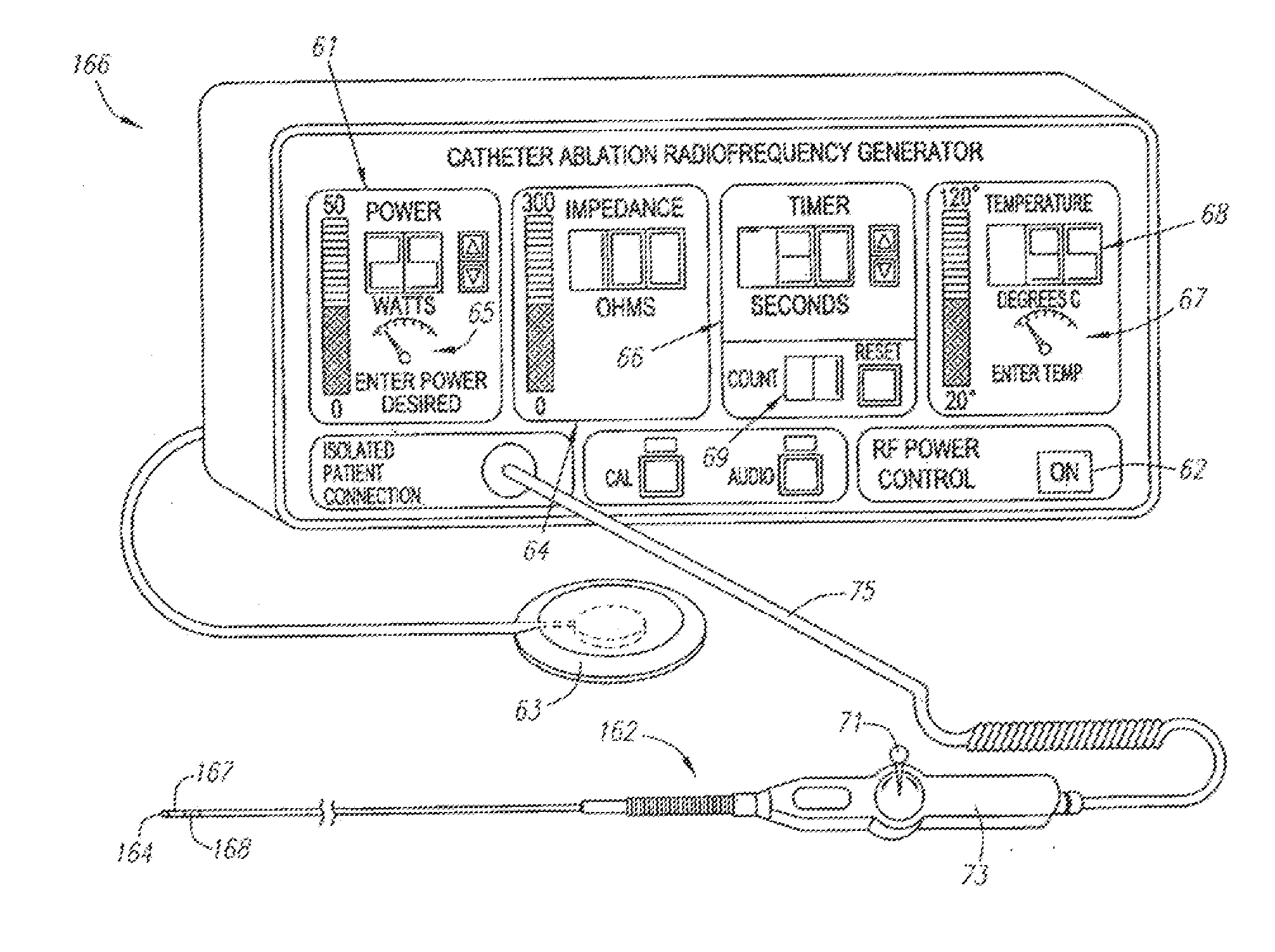
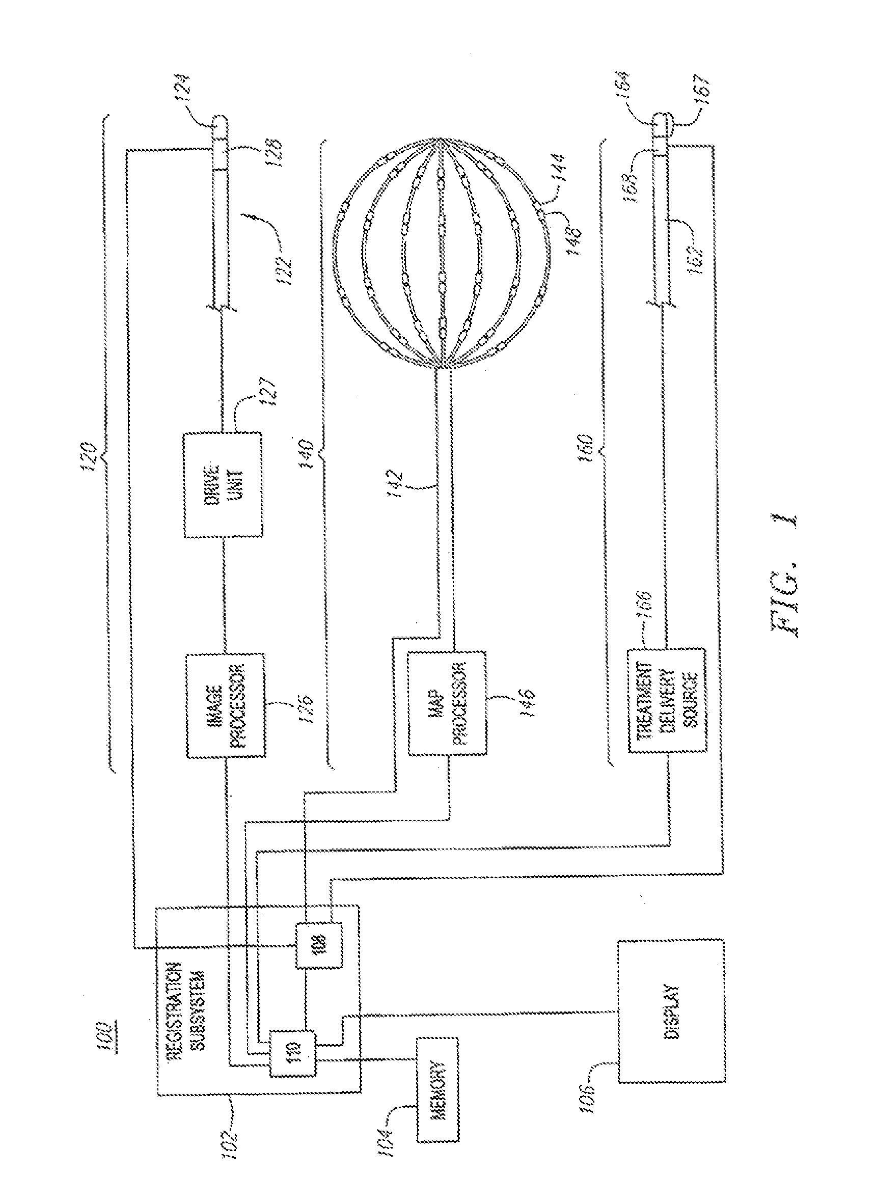
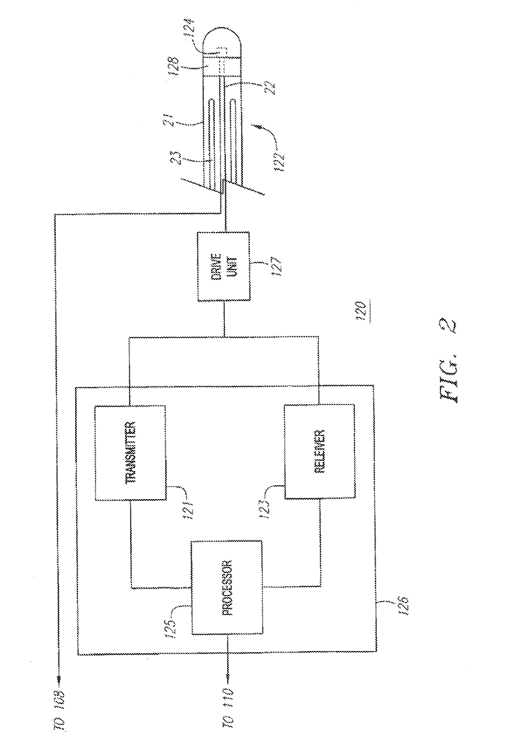
[0036]The present invention provides a system for generating a three-dimensional image of a volume, registering that image in a three-dimensional coordinate system, generating mapping data of the volume, registering the positional data to the three-dimensional coordinate system, and guiding a treatment device to a target site identified by the positional data. The system is particularly suited for reconstructing and mapping a volume within a heart, and for ablating heart tissue. Nevertheless, it should be appreciated that the invention is applicable for use in other applications. For example, the various aspects of the invention have application in procedures for ablating or otherwise treating tissue in the prostate, brain, gall bladder, uterus, esophagus and other regions of the body. Additionally, it should be appreciated that the invention is applicable for use in drug therapy applications where a therapeutic agent is delivered to a targeted tissue region. One preferred embodimen...


PUM


Abstract

Systems and methods for imaging a body cavity and for guiding a treatment element within a body cavity are provided. A system may include an imaging subsystem having an imaging device and an image processor that gather image data for the body cavity. A mapping subsystem may be provided, including a mapping device and a map processor, to identify target sites within the body cavity, and provide location data for the sites. The system may also include a location processor coupled to a location element on a treatment device to track the location of the location element. The location of a treatment element is determined by reference to the location element. A treatment subsystem including a treatment device having a treatment element and a treatment delivery source may also be provided. A registration subsystem receives and registers data from the other subsystems, and displays the data.

Description

RELATED APPLICATIONS[0001]The present application is a continuation-in-part of copending U.S. application Ser. No. 10 / 012,293.FIELD OF THE INVENTION[0002]The present inventions relate generally to systems and methods for guiding and locating diagnostic or therapeutic elements on medical instruments positioned in a body.BACKGROUND[0003]The use of invasive medical devices, such as catheters and laparoscopes in order to gain access into interior regions or volumes within the body for performing diagnostic and therapeutic procedures is well known. In such procedures, it is important for a physician or technician to be able to precisely position the device, including various functional elements located on the device, within the body in order to make contact with a desired body tissue location.[0004]In order to accurately position the device, it is desirable that the shape or configuration of the particular volume be determined, and registered in a known three-dimensional coordinate syste...

Claims


Application Information

Patent Timeline
Patent Type & Authority Applications(United States)
IPC IPC(8): A61B6/03A61B8/12A61B5/055A61B6/12A61B5/042A61B5/06A61B17/00A61B18/14A61B19/00G06T7/00G06T17/40
CPCA61B5/0422G06T2210/41A61B5/6858A61B8/543A61B18/1492A61B19/52A61B19/5244A61B2017/00053A61B2017/00703A61B2018/00214A61B2018/00267A61B2018/00357A61B2018/00839A61B2019/505A61B2019/5251A61B2019/5253A61B2019/5263A61B2019/5272A61B2019/5276A61B2019/5278A61B2019/5291A61B2019/5429A61B2019/5458A61B2562/0219G06T7/0024G06T19/003G06T19/006G06T2207/30048A61B5/06A61B90/36A61B2034/2053A61B2034/2063A61B2034/2072A61B2090/3782A61B34/20A61B2034/105A61B2034/2051A61B2090/378A61B2090/365A61B2090/3929A61B2090/3958G06T7/30A61B5/062A61B5/287
Inventor PANESCU, DORIN
Owner BOSTON SCI SCIMED INC
Why Eureka
Try Eureka