Radiotherapy dose assessment and adaption using online imaging

Inactive Publication Date: 2016-09-29
SONITRACK SYST
View PDF2 Cites 34 Cited by
  • Summary
  • Abstract
  • Description
  • Claims
  • Application Information

AI Technical Summary

Benefits of technology

[0006]The deformed planning scans can then be used to compute radiation delivered to the patient in a manner that better represents the state of the patient's actual anatomy during beam delivery. While radiotherapy treatment is described, such methods are not limited to radiotherapy but can utilize a number of other medical therapies where the treatment dose can be planned and assessed, including but not limited to, high intensity focused ultrasound therapy (HIFU), radiofrequency ablations, hypothermic therapies, hyperthermic therapies, etc.
[0007]One method for estimating dose delivered during medical therapy delivery may comprise acquiring one or more planning scans of a portion of a patient body prior to medical therapy delivery; acquiring one or more online images of the portion of the patient body or in proximity to the portion prior to or during medical therapy delivery; deforming the one or more planning scans in accordance with a presentation of the one or more online images to create one or more deformed planning scans; and estimating a dose for delivery to the portion of the patient body during the medical therapy delivery using the one or more deformed planning scans. The one or more online images do riot need to align directly with or correspond to the one of more planning scans;

Problems solved by technology

However, since the planning scan is obtained prior to treatment, (potentially days or weeks prior), it does not necessarily represent the state of the patient's anatomy as it presents at the time of treatment beam delivery.
The potential mismatch between the patient's anatomy in

Method used


Image

  • Radiotherapy dose assessment and adaption using online imaging
  • Radiotherapy dose assessment and adaption using online imaging
  • Radiotherapy dose assessment and adaption using online imaging

Examples


Embodiment Construction

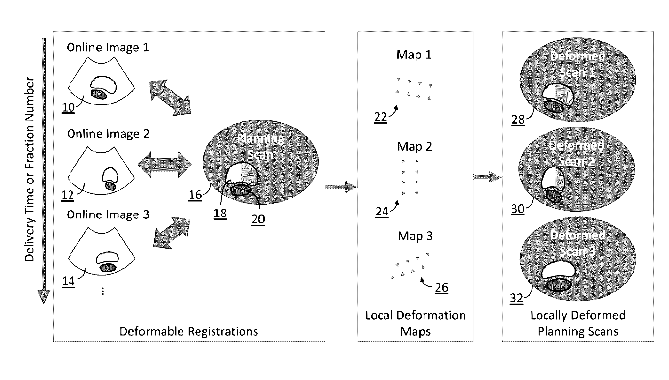
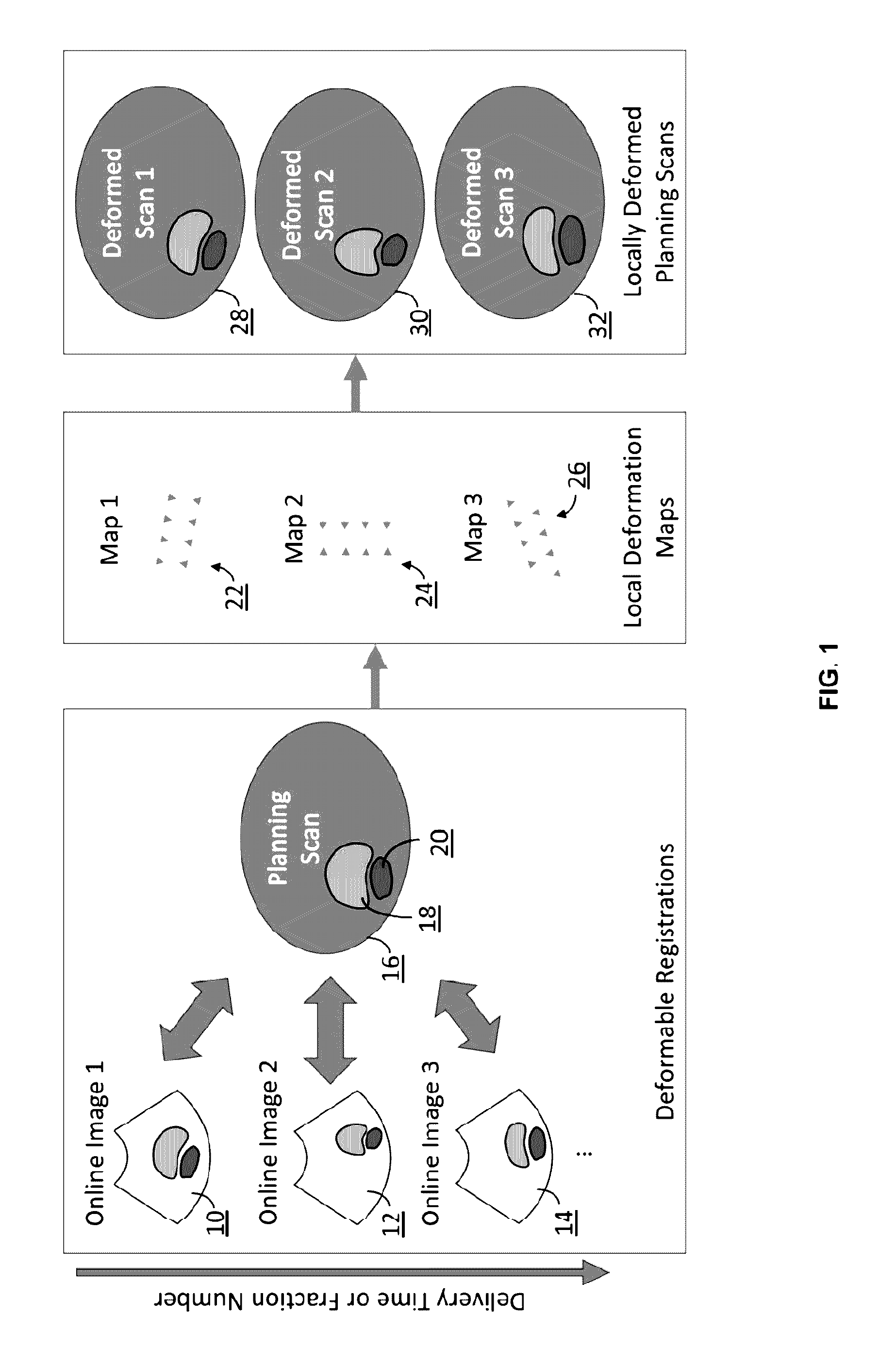
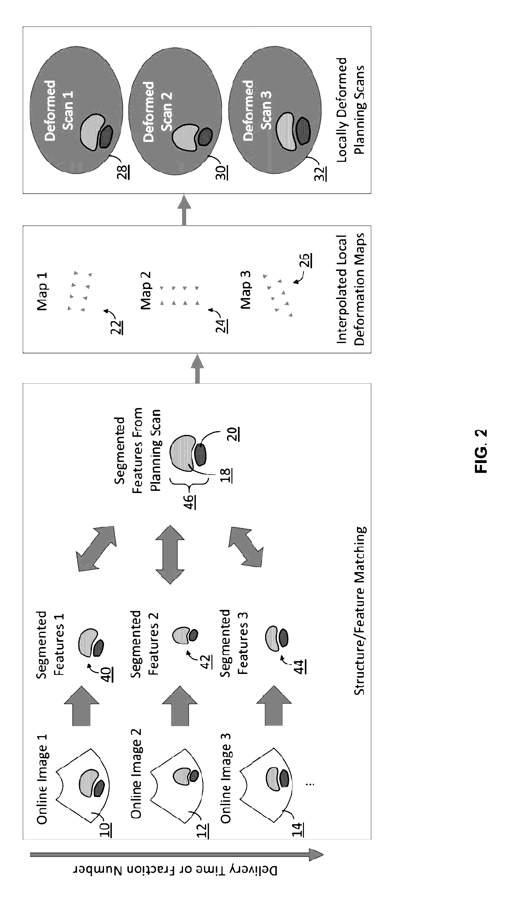
[0019]The methods described herein use information from online imaging scans collected before and / or during radiation therapy beam delivery in order to assess and adapt radiation dose delivered to the patient. The online images capture the state of the patient's anatomy directly prior to or during radiation beam delivery. The premise is to use the online images to inform deformations to the planning scans that were originally used to plan and simulate the radiation dose delivered to the patient. The deformed planning scans can then be used to compute radiation delivered to the patient in a mariner that better represents the state of the patient's actual anatomy during beam delivery. Note that while the methods below are discussed in the context of radiotherapy, it is also possible to apply such methods to other areas of medical therapy where dose can be planned and assessed including but not limited to high intensity focused ultrasound therapy (HIFU), radiofrequency ablations, hypot...


PUM


Abstract

In external beam radiation therapy, a planning image (scan) of the patient is obtained prior to treatment as a basis for constructing a radiation delivery plan. However, since the planning scan is obtained prior to treatment (potentially days or weeks prior), it does not necessarily represent the state of the patient's anatomy as it presents at the time of treatment beam delivery. The potential mismatch between the patient's anatomy in the planning scan and anatomy at the time of treatment can result in dose discrepancies between the planned dose and the actual delivered dose. The methods herein describe the use of online images taken immediately before or during treatment delivery in order to predict, assess, and adapt to such discrepancies.

Description

CROSS-REFERENCE TO RELATED APPLICATIONS[0001]This application is a continuation of International Application No. PCT / US2014 / 068927 filed Dec. 5, 2014, which claims the benefit of priority to U.S. Provisional Patent Application No. 61 / 912,985 filed Dec. 6, 2013, each of which is incorporated herein by reference in its entirety.FIELD OF THE INVENTION[0002]The present invention relates to methods and apparatus for monitoring, predicting, and adapting radiation doses based on imaging patients immediately prior to and / or during radiation beam delivery.BACKGROUND OF THE INVENTION[0003]External Beam Radiation Therapy (EBRT) is used to treat more than half of all cancer patients worldwide. Traditionally in EBRT, a planning image (scan) of the patient (usually a CT or MRI image) is obtained prior to treatment as a basis for constructing a radiation delivery plan including beam angles, shapes, and intensities. The delivery plan is simulated using the information in the planning scan in order ...

Claims


Application Information

Patent Timeline
IPC IPC(8): A61N5/10
CPCA61N5/1071A61N5/1039A61N2005/1058A61N5/1049A61N2005/1055A61N5/1045A61N5/107A61N2005/1072A61N7/02A61B18/12A61B2034/104
Inventor SCHLOSSER, JEFFREY
Owner SONITRACK SYST
Why Eureka
Try Eureka