Visualizing Radiation Therapy Beam in Real-Time in the context of Patient's Anatomy

a radiation therapy and anatomy technology, applied in the field of radiation therapy, can solve the problems of unacceptable risk for patients and healthcare providers, underdosing or overdosing patients, and affecting the patient's anatomy

Active Publication Date: 2015-12-17
THE BOARD OF TRUSTEES OF THE LELAND STANFORD JUNIOR UNIV
View PDF2 Cites 39 Cited by
  • Summary
  • Abstract
  • Description
  • Claims
  • Application Information

AI Technical Summary

Benefits of technology

[0022]According to another embodiment of the invention, a method of radiation therapy quality assurance assessment is provided that includes coating a phantom with a scintillating material that conforms to the external shape of the phantom, and irradiating the coated phantom, using a therapeutic beam from a medical source of ionizing radiation, where the scintillating material emits light when exposed to the source of ionizing radiation, where the emitted light is collected by a camera, where the camera forms image data, where the image data is sent to an appropriately programmed computer, where the data is processed to determine characteristics of the beam and to determine an accuracy of machine operation for output to an operator.
[0023]According to one aspect of the current embodiment, the collected image data is used to determine acceleration, velocity, accuracy and repeatability of mechanical motions of the source of ionizing radiation. In one aspect, the medical source of ionizing radiation includes a linear accelerator where evaluated mechanical motions are selected from the group consisting of motions of the gantry, collimator, collimators, source and couch. In a further aspect, the source of radiation comprises a radioactive seed, where the evaluated motions include the motions of the speed.

Problems solved by technology

Although most RT treatments are performed accurately, accidents from equipment faults, failures in quality assurance methodologies, and errors during operation represent an unacceptable risk for both patients and healthcare providers.
However, these numbers may be grossly underestimated, as it is also widely recognized that many incidents remain unreported during RT.
A significant number of these incidents occur during the delivery of RT and can result in underdosing or overdosing patients, irradiating healthy tissue, or, at worst, patient death.
While numerous methods [e.g., in room lasers, tattoos on patient skin, mega- and kilovoltage (MV, kV) imaging, pretreatment dosimetry phantom studies, etc.] have been developed to ensure the fidelity of treatment delivery, there are no technologies currently available that are capable of fully monitoring treatment in the context of the patient's anatomy as it is being delivered.
However, due to a lack of soft tissue contrast and multileaf collimator (MLC) blockage of the field of view, the available anatomic references provided by the approach are limited to bony anatomy or implanted fiducial markers that lie within the beam's eye view making the interpretation of the data nontrivial.
Thus, these techniques require long exposure times in darkened rooms or complex, expensive imaging setups to obtain usable measurements.
This poses a challenge to practical application.
Therefore, the ability to visualize the position, shape, and intensity of the radiation beam as it passes through the patient in real time is not yet available in clinical practice.
Additionally, in the radiation therapy space, a great deal of effort is expended upon ensuring that medical sources of ionizing radiation operate properly.
Often these tests are time-consuming, tedious, and prone to operator error.

Method used


Image

  • Visualizing Radiation Therapy Beam in Real-Time in the context of Patient's Anatomy
  • Visualizing Radiation Therapy Beam in Real-Time in the context of Patient's Anatomy
  • Visualizing Radiation Therapy Beam in Real-Time in the context of Patient's Anatomy

Examples


Embodiment Construction

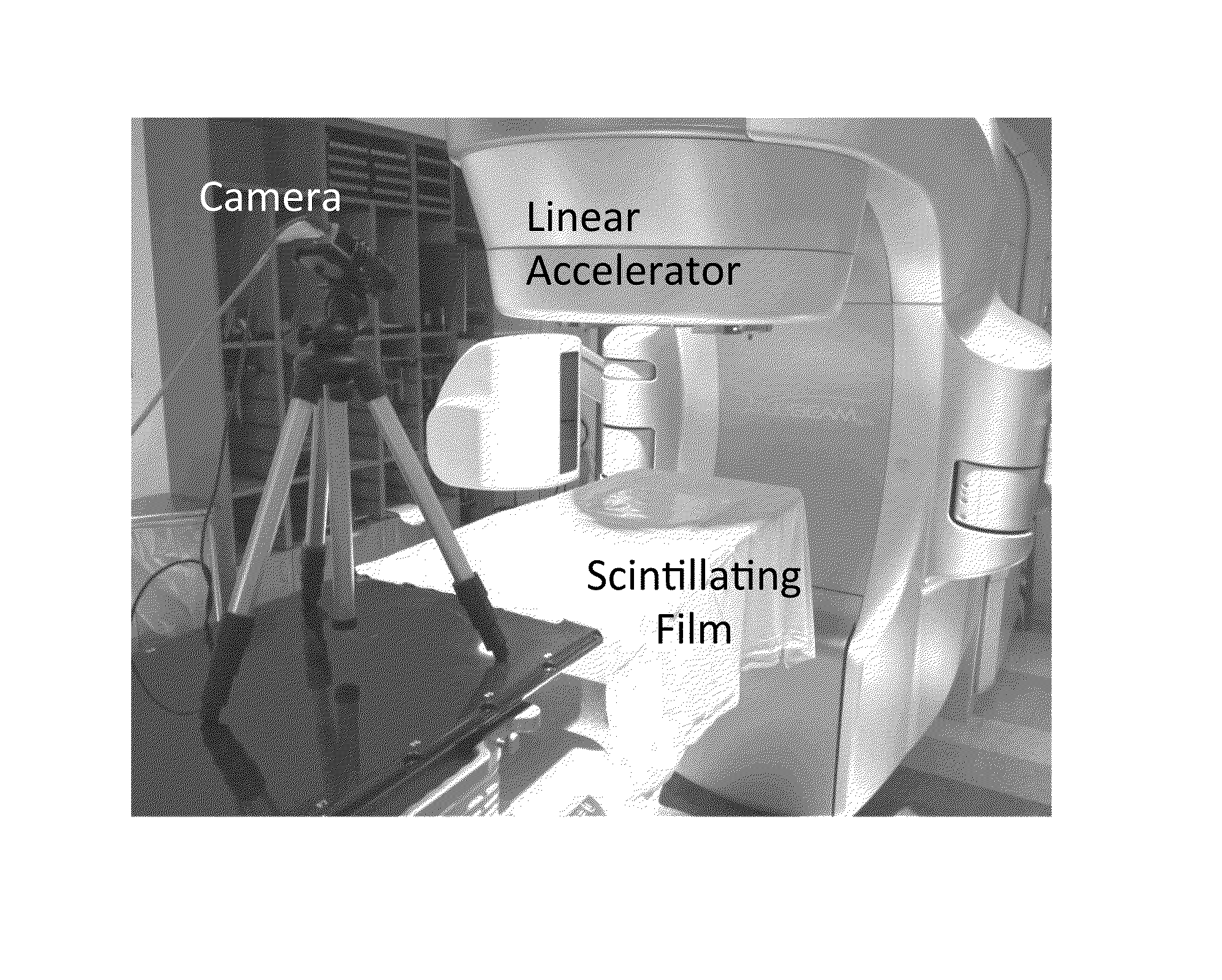
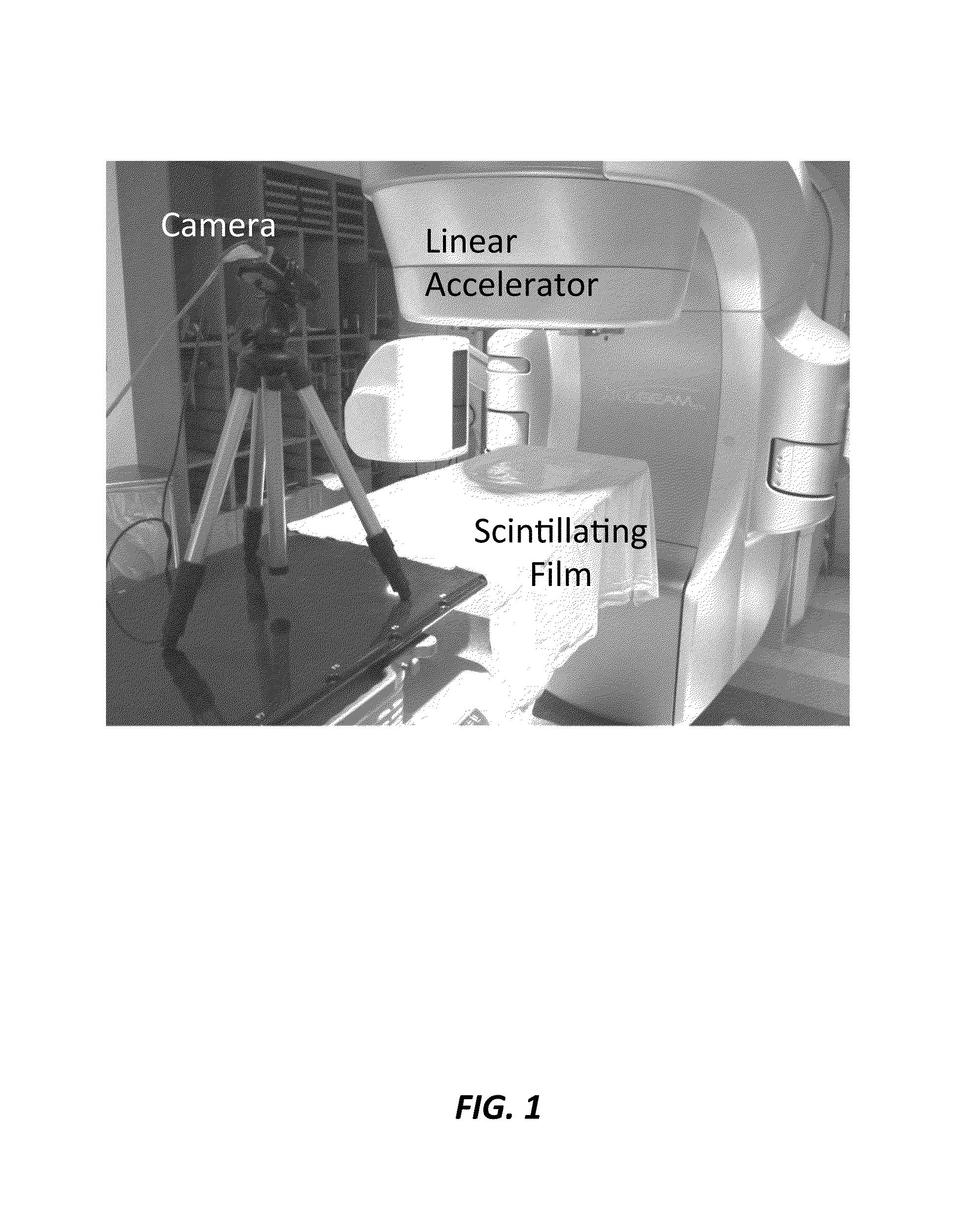
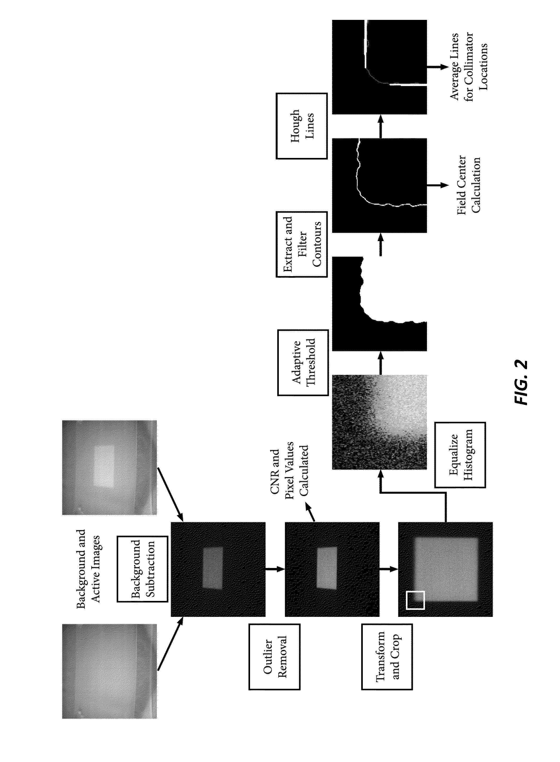
[0040]The current invention is a real-time beam visualization (RT-BV) system and method that includes of a flexible scintillating film and a camera. In one embodiment, the film is placed on a patient's skin such that it will emit an optical signal when the beam passes through it. The camera is arranged such that it can image the emitted signal as well as the surrounding patient surface anatomy. An exemplary embodiment is provided herein that includes characterization and integration of the RT-BV system for application in a RT treatment room. An image-processing algorithm was developed to demonstrate how the data from the RT-BV system could be analyzed and interpreted to provide quantitative treatment verification, as well as to aid in assessing the quality of the data from the system.

[0041]According to one embodiment, a method of real-time radiotherapy beam visualization is provided that includes disposing a free-form flexible scintillating sheet on a subject of interest, irradiatin...


PUM


Abstract

A method of real-time radiotherapy beam visualization is provided that includes disposing a free-form flexible scintillating sheet on a subject of interest, irradiating the subject of interest with a source of ionizing radiation, where the free-forming flexible scintillating sheet emits light when irradiated by the therapeutic photon beam, collecting the emitted light and collecting ambient light reflected from the subject of interest and surrounding objects using a camera, where the collected light is converted to image data by the camera, where the image data is communicated to an appropriately programmed computer, and processing the image data to determine beam characteristics and the characteristics of the subject of interest, using the appropriately programmed computer, where the beam characteristics and the characteristics of the subject of interest are displayed in real-time to a machine operator enabling real-time verification of treatment delivery.

Description

CROSS-REFERENCE TO RELATED APPLICATIONS[0001]This application claims priority from U.S. Provisional Patent Application 62 / 012,710 filed Jun. 16, 2014, which is incorporated herein by reference.FIELD OF THE INVENTION[0002]The present invention relates generally to radiotherapy. More particularly, the invention relates to a system and method of visualizing a radiotherapy treatment beam entrance or exit relative to the patient.BACKGROUND OF THE INVENTION[0003]External beam radiation therapy (EBRT) is one of the most commonly used treatments for numerous cancer types. In 2010, approximately 470,000 patients underwent radiation therapy (RT) in the US alone. Depending on their individual treatment plan, these patients may undergo between 1 and 70 separate RT visits over the course of several weeks. Although most RT treatments are performed accurately, accidents from equipment faults, failures in quality assurance methodologies, and errors during operation represent an unacceptable risk fo...

Claims


Application Information

Patent Timeline
Patent Type & Authority Applications(United States)
IPC IPC(8): A61N5/10
CPCA61N2005/1076A61N5/1075A61N5/1049A61N2005/1059
Inventor XING, LEINACZYNSKI, DOMINIK JANJENKINS, CESARE
Owner THE BOARD OF TRUSTEES OF THE LELAND STANFORD JUNIOR UNIV
Why Eureka
Try Eureka