

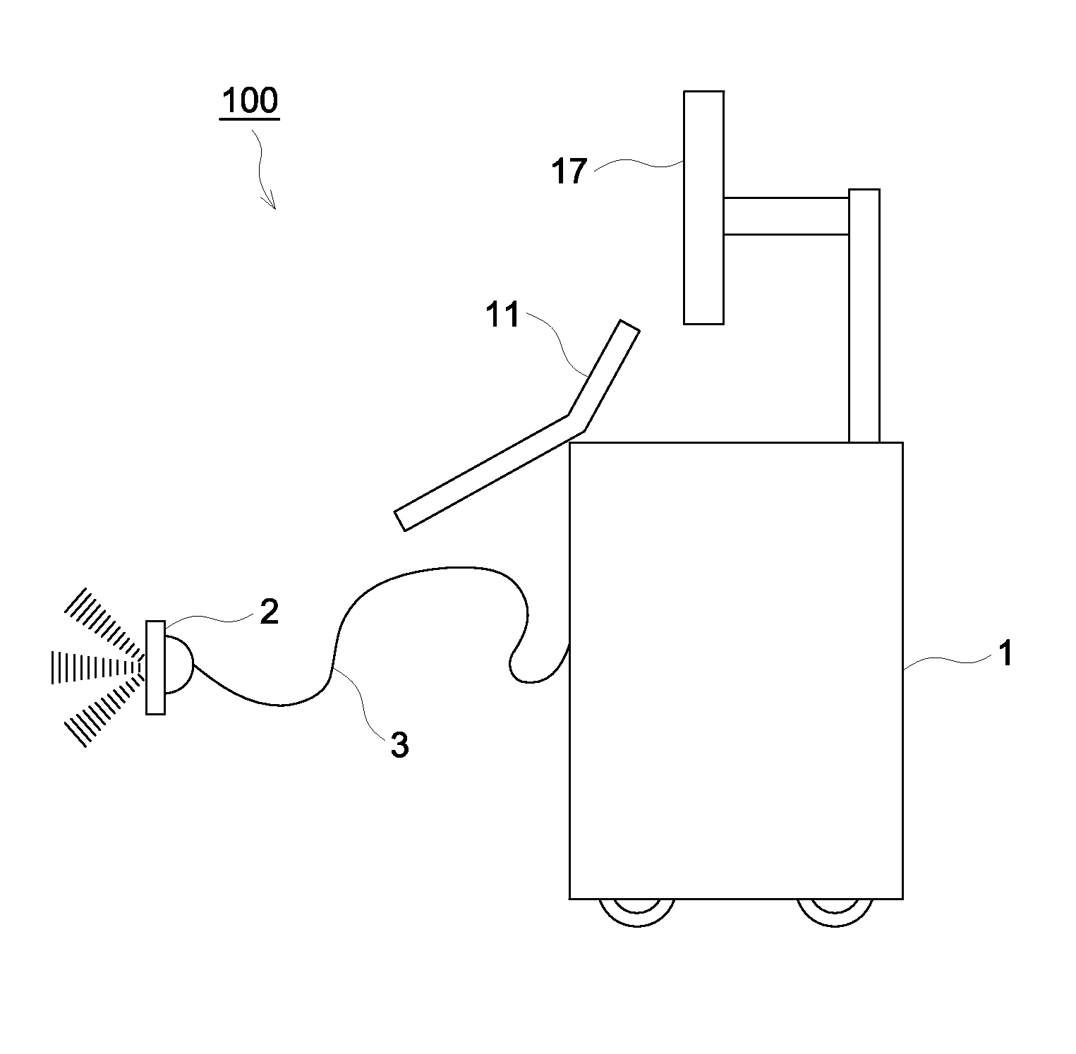
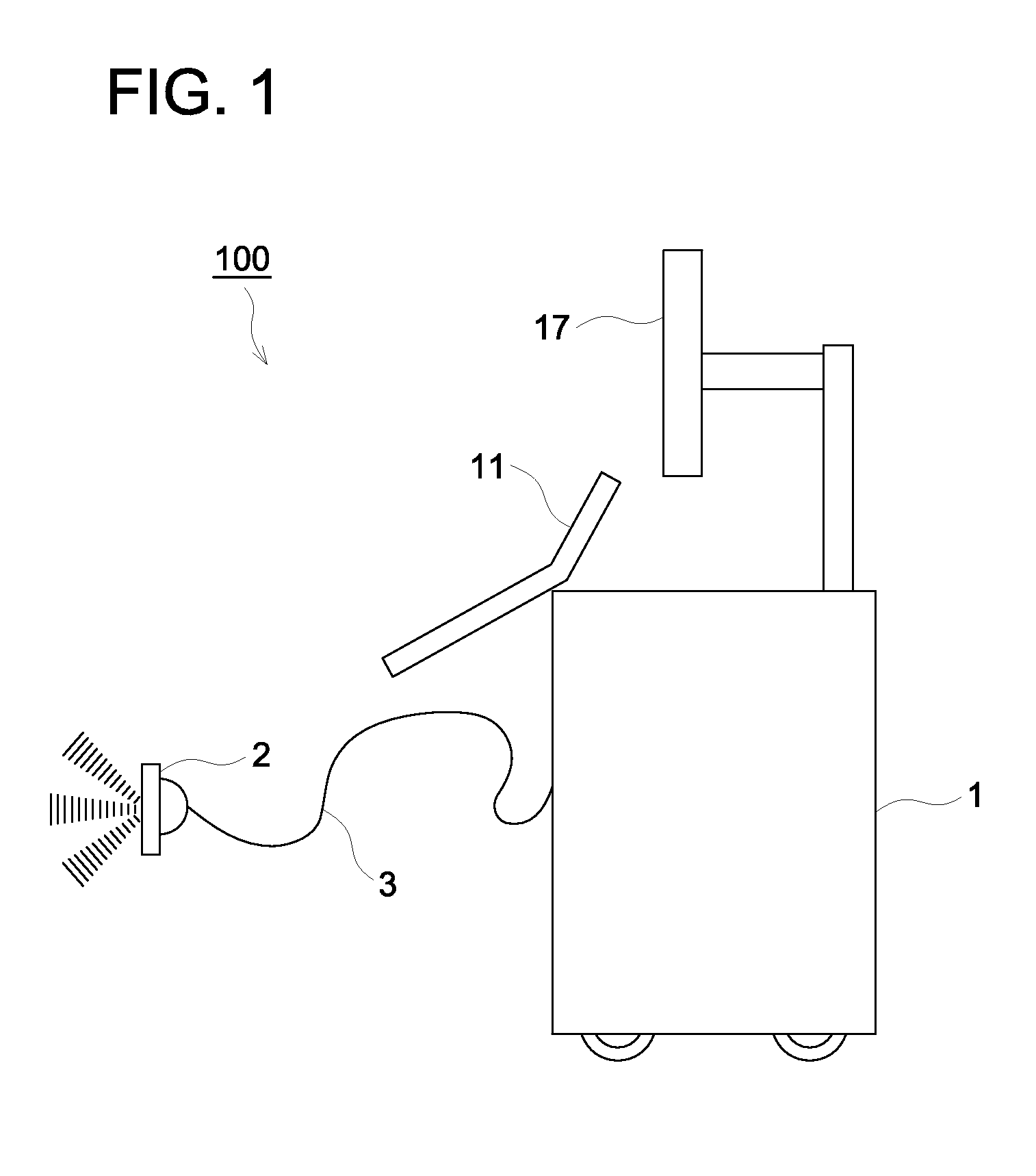
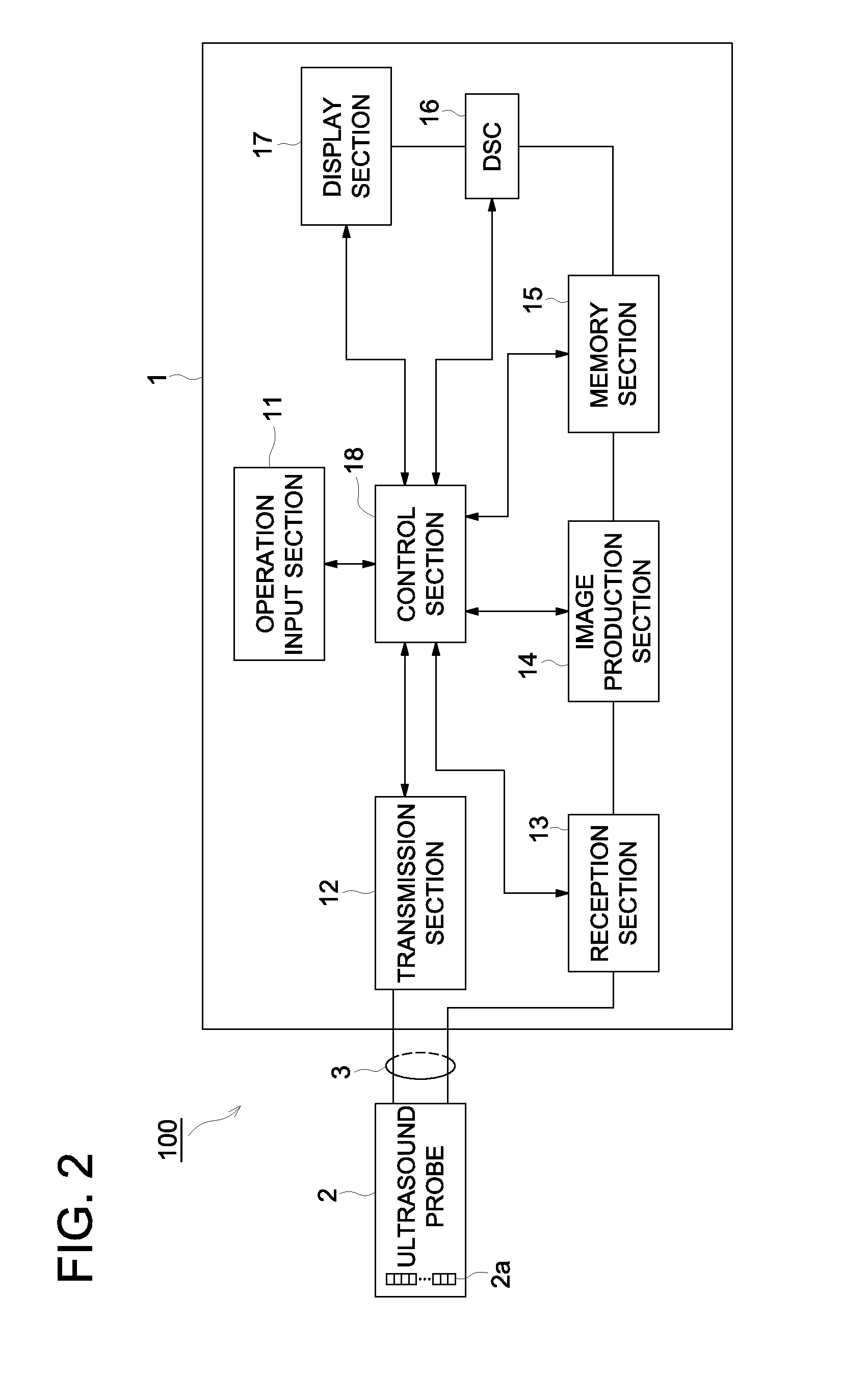
Ultrasound diagnostic device
a diagnostic device and ultrasound technology, applied in tomography, instruments, applications, etc., can solve the problems of not always subjecting all signals to correct beam forming, the actual delay amount may differ from the theoretical value, and the acoustic velocity of ultrasound in a tested subject is not always constan
- Summary
- Abstract
- Description
- Claims
- Application Information
AI Technical Summary
Benefits of technology
Problems solved by technology
Method used
Image

Examples
example 1
[0075]A reception signal having been obtained via transmission / reception of ultrasound with respect to a given phantom having a plurality of wires using the ultrasound diagnostic device 100 according to the present embodiment was subjected to beam forming. A reception signal after beam forming was multiplied by a coherence factor set at −18 dB as the lower limit to be corrected, on the basis of which imaging was carried out. The image is shown in FIG. 7.
example 2
[0076]In the same manner, the lower limit was set at −18 dB and then a reception signal after beam forming was multiplied by a corrected coherence factor having been obtained via exponentiation of a correction value (γ=0.75) to be corrected, on the basis of which imaging was carried out. The image is shown in FIG. 8.
example 3
[0077]In the same manner, the lower limit was set at −18 dB and then a reception signal after beam forming was multiplied by a corrected coherence factor having been obtained via exponentiation of a correction value (γ=0.5) to be corrected, on the basis of which imaging was carried out. The image is shown in FIG. 9.
PUM
 Abstract
Description
Claims
Application Information