Biological Pacemaker

a pacemaker and biotechnology, applied in the direction of viruses, drug compositions, cardiovascular disorders, etc., can solve the problems of significant cost, infection, lung collapse, and inability to fully realize the effect of ventricular valve stimulation

Inactive Publication Date: 2012-12-27
THE JOHN HOPKINS UNIV SCHOOL OF MEDICINE
View PDF0 Cites 2 Cited by
  • Summary
  • Abstract
  • Description
  • Claims
  • Application Information

AI Technical Summary

Benefits of technology

[0012]We now provide gene transfer and cell administration methods that can create a pacemaker function, and / or modulate the activity of an endogenous or induced cardiac pacemaker function.
[0022]The administered polynucleotide suitably induces or modulates (increase or decrease) at least one heart electrical property. Preferred use of the invention modulates heart electrical conduction and preferably can reconfigure all or part of the cardiac action potential (AP). That use helps achieve a desired therapeutic outcome. Significant disruption of normal electrical function is usually reduced and often avoided by the present methods.

Problems solved by technology

However there has been long-standing recognition that abnormalities of excitable cardiac tissue can lead to abnormalities of the heart rhythm.
However, such implanted devices have significant inherent risks such as infection, hemorrhage, lung collapse as well as significant expense.

Method used


Image

  • Biological Pacemaker
  • Biological Pacemaker
  • Biological Pacemaker

Examples


example 1

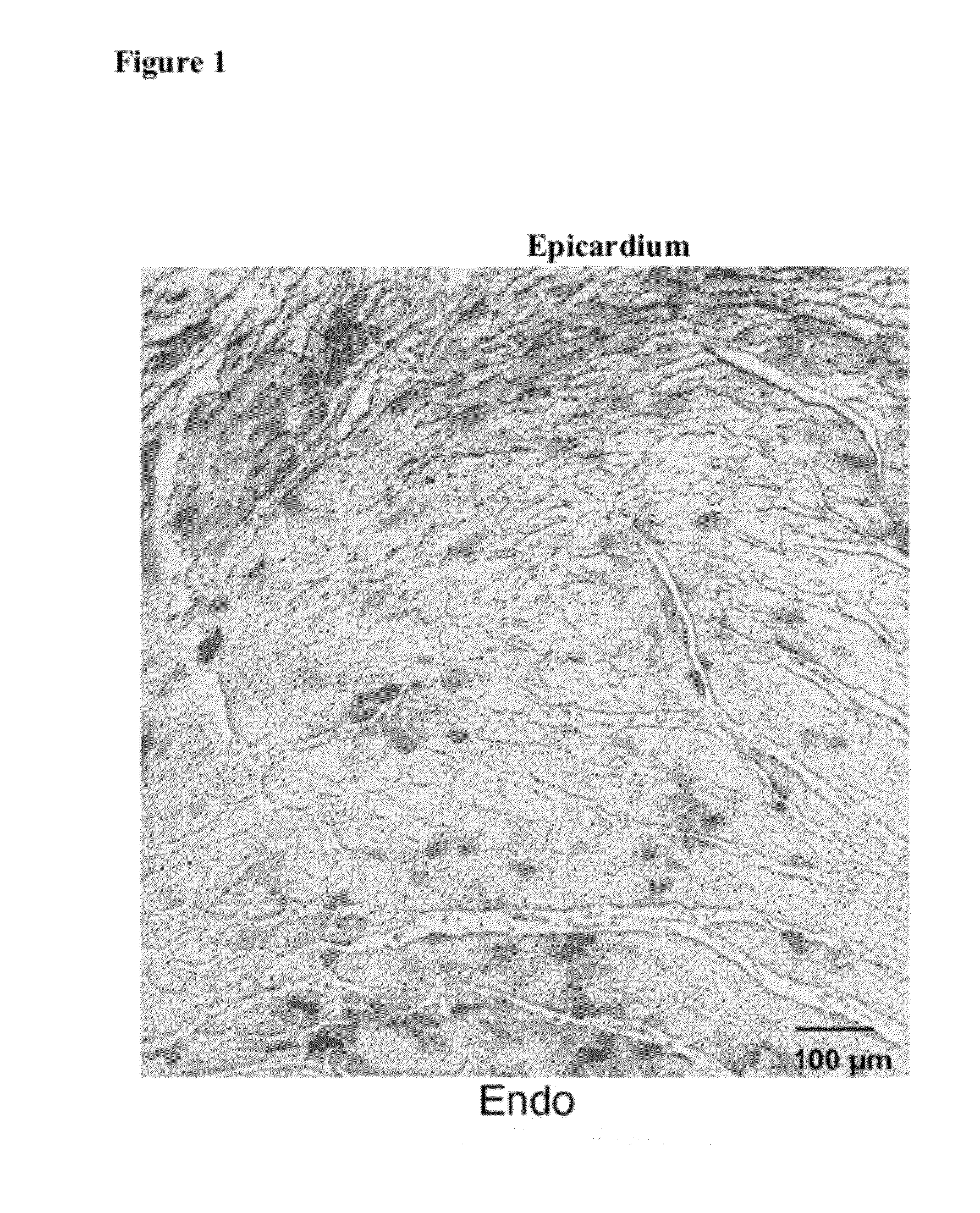
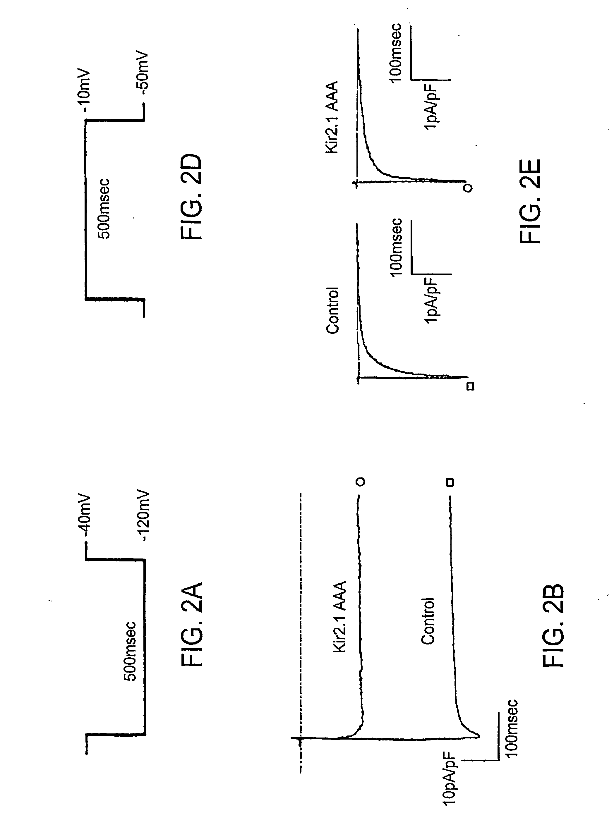
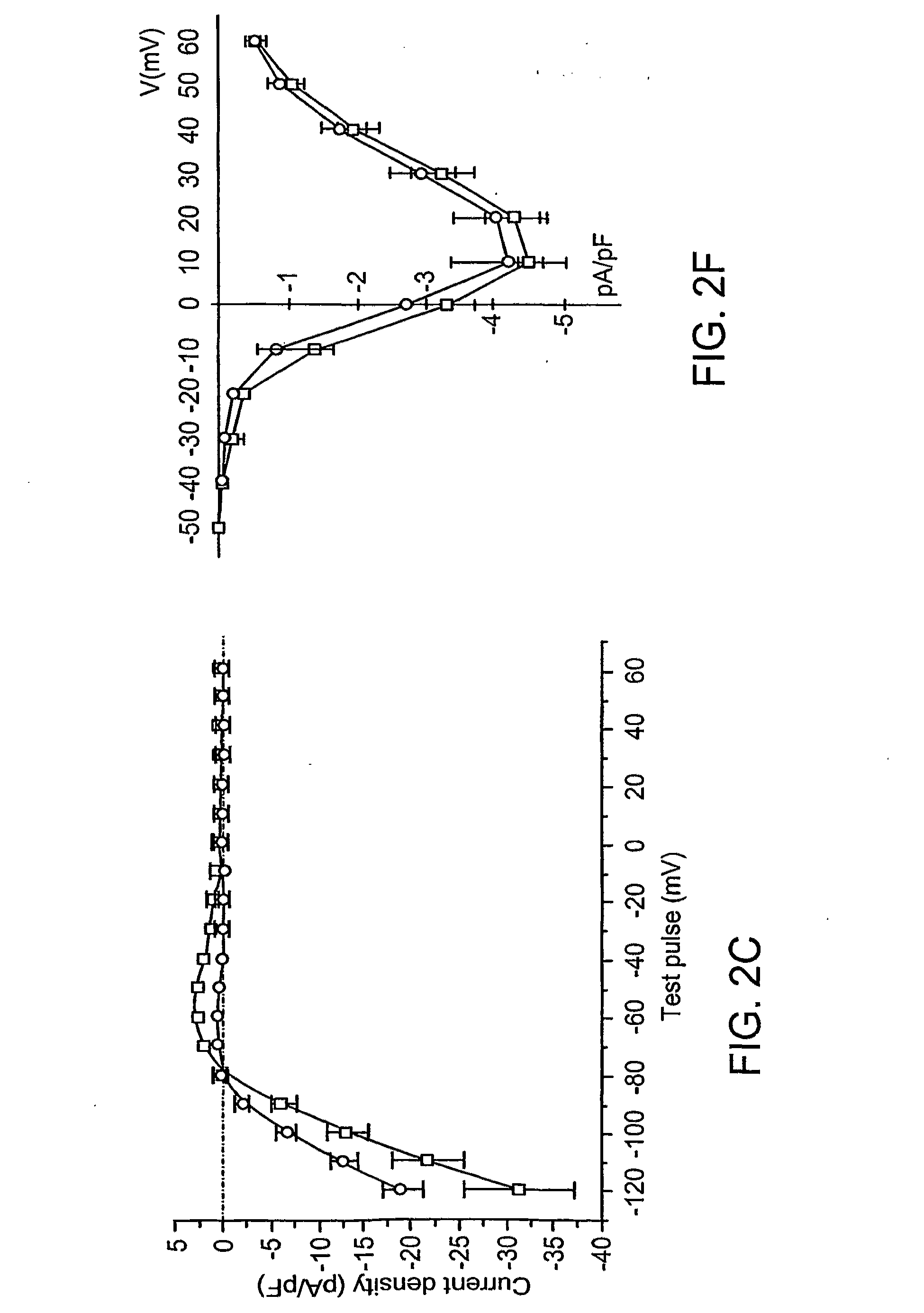
Effect of Inhibition of Kir2 Channels on Latent Pacemaker Activity of Ventricular Myocytes

Materials and Methods

[0131]Dominant-Negative Effects

[0132]Dominant-negative effects of Kir2.1AAA on IK1 expression were achieved using the approach of Herskowitz (See, for example I. Herskowitz, Nature 329, 219-22; (1987)). The GYG motif, three amino acids in the H5 region of potassium channels that play a key role in selectivity and pore function, were replaced with three alanines in Kir2.1.

[0133]Vectors

[0134]A bicistronic adenoviral vector, encoding both enhanced green fluorescence protein (EGFP, Clontech, Palo Alto, Calif., USA) and Kir2.1AAA, was created using the adenovirus shuttle vectors pAdEGI (D. C. Johns, H. B. Nuss, E. Marban, J. Biol. Chem. 272, 31598-603. (1997) and pAdC-DBEcR (U. C. Hoppe, E. Marban, D. C. Johns, J. Clin. Invest. 105, 1077-84. (2000)) as previously described— The full-length coding sequence of human Kir2.1 (kindly supplied by G.F. Tomaselli, Johns Hopkins Universi...

example 2

The Triple Mutation GYG365-367AAA Rendered HCN1 Channels Nonfunctional

Molecular Biology and Heterologous Expression

[0150]mHCN1 and mHCN2 were subcloned into the pGH expression vector. B. Santoro et al., Cell, 93:717-29 (1998). Site-directed mutagenesis was performed using polymerase chain reaction (PCR) with overlapping mutagenic primers. All constructs were sequenced to ensure that the desired mutations were present. cRNA was transcribed from NheI- and SphI-linearized DNA using T7 RNA polymerase (Promega, Madison, Wis.) for HCN1 and HCN2 channels, respectively. Channel constructs were heterologously expressed and studied in Xenopus oocytes. Briefly, stage IV through VI oocytes were surgically removed from female frogs anesthetized by immersion in 1% tricaine (i.e. 3-aminobenzoic acid ethyl ester) followed by digestion with 2 mg / mL collagenase in OR-2 containing (in mM): 88 NaCl, 2 KCl, 1 MgCl2 and 5 mM HEPES (pH 7.6 with NaOH) for 30 to 60 minutes. Isolated oocytes were injected wi...


PUM

PropertyMeasurementUnit
membrane potentialsaaaaaaaaaa

Abstract

Disclosed are methods and systems for modulating electrical behavior of cardiac cells. Preferred methods include administering a polynucleotide or cell-based composition that can modulate cardiac contraction to desired levels, i.e., the administered composition functions as a biological pacemaker.

Description

[0001]The present application claims priority to U.S. Provisional Application No. 60 / 287,088 filed on Apr. 27, 2001, which is incorporated by reference herein in its entirety.STATEMENT OF FEDERALLY SPONSORED RESEARCH[0002]Funding for the present invention was provided in part by the Government of the United States by virtue of Grant No. NIH P50 HL52307 by the National Institutes of Health. Thus, the Government of the United States has certain rights in and to the invention claimed herein.BACKGROUND[0003]1. Field of the Invention[0004]The invention generally features methods to provide and / or modulate a cardiac pacemaker function. In preferred aspects, the invention provides genetically-engineered pacemakers that can be employed as an alternative or supplement to implantable electronic pacemakers to induce or modulate ventricular or atrial firing rate.[0005]2. Background[0006]Spontaneous cellular electrical rhythms govern numerous biological processes from the autonomous beating of t...

Claims


Application Information

Patent Timeline
Patent Type & Authority Applications(United States)
IPC IPC(8): A61K31/7088C12N15/85A61P9/00C12N5/10A61K35/00A61P9/04C12N15/09A61K38/00A61K48/00A61P9/06C07K14/705C12Q1/02C12Q1/68
CPCA61K38/00C12N15/85C12N2799/022C07K14/705A61P9/00A61P9/04A61P9/06
Inventor MARBAN, EDUARDOLI, RONALD
Owner THE JOHN HOPKINS UNIV SCHOOL OF MEDICINE
Why Eureka
Try Eureka