Method and apparatus for processing medical image, and robotic surgery system using image guidance

a robotic surgery and medical image technology, applied in tomography, instruments, applications, etc., can solve the problems of difficult for surgeons to perceive the correct position and shape of the part to be operated, and the effect of affecting the operation efficiency of the surgery

Inactive Publication Date: 2013-02-07
SAMSUNG ELECTRONICS CO LTD
View PDF14 Cites 136 Cited by
  • Summary
  • Abstract
  • Description
  • Claims
  • Application Information

AI Technical Summary

Benefits of technology

[0010]According to one or more other embodiments, an apparatus for processing a medical image includes: an image acquisition unit for acquiring medical images captured using a plurality of multi-modal medical image capturing apparatuses with respect to a predetermined organ; a surface information extractor for extracting surface information of the predetermined organ, which is contained in each of the medical images, from each of the medical images; an image mapping unit for mapping the medical images by using the extracted surface information; and a synthesis image generator for generating a synthesis image in which the medical images have been registered, based on the mapping result.
[0011]According to one or more other embodiments, a robotic surgery system for performing a robotic surgery by a surgery robot by guiding images of a part to be operated includes: an endoscope apparatus for capturing medical images of a predetermined organ in a body to be examined; a non-endoscopic apparatus including at least one of an ultrasound apparatus, a computed tomography (CT) apparatus, a magnetic resonance imaging (MRI) apparatus, and a positron emission tomography (PET) apparatus for capturing medical images of the predetermined organ; a medical image processing apparatus for acquiring the medical images captured using the plurality of multi-modal medical image capturing apparatuses, extracting surface information of the predetermined organ, which is contained in each of the medical images, from each of the medical images, mapping the medical images by using the extracted surface information, and generating a synthesis image in which the medical images have been registered, based on the mapping result; a display apparatus for displaying the generated synthesis image; and the surgery robot for performing a robotic surgery according to a user input.
[0012]According to one or more other embodiments, a method of processing a medical image includes acquiring first image data of a predetermined organ in a living body using a first medical image capturing apparatus and second image data of the predetermined organ using a second medical image capturing apparatus that is different than the first, extracting first surface information of the predetermined organ from the first image data and second surface information of the predetermined organ from the second image data, matching a first position obtained from the first surface information to a second position obtained from the second surface information in accordance with relative position information of the first and second medical image capturing apparatuses, wherein the first position corresponds to the second position, generating a synthesis image from the first surface information and the second surface information based on the matching and outputting or displaying the synthesis image.

Problems solved by technology

While surgeons perform robotic surgeries after having perceived a part to be operated on using computed tomography (CT) images, magnetic resonance imaging (MRI) images, and ultrasound images, there is a limitation in that the robotic surgeries significantly depend on the experience of the surgeons.
However, because images of a part to be operated on that are acquired with an endoscope, such as a laparoscope, are only images of surfaces of a patient's internal organs, when the part to be operated is not viewed by the scope because it is hidden by other organs or because it is inside other organs, it is difficult for a surgeon to perceive a correct position and shape of the part to be operated on.

Method used


Image

  • Method and apparatus for processing medical image, and robotic surgery system using image guidance
  • Method and apparatus for processing medical image, and robotic surgery system using image guidance
  • Method and apparatus for processing medical image, and robotic surgery system using image guidance

Examples


Embodiment Construction

[0027]Reference will now be made in detail to embodiments, examples of which are illustrated in the accompanying drawings, wherein like reference numerals refer to like elements throughout. In this regard, the present embodiments may have different forms and should not be construed as being limited to the descriptions set forth herein. Accordingly, the embodiments are merely described below, by referring to the figures, to explain aspects of the present description.

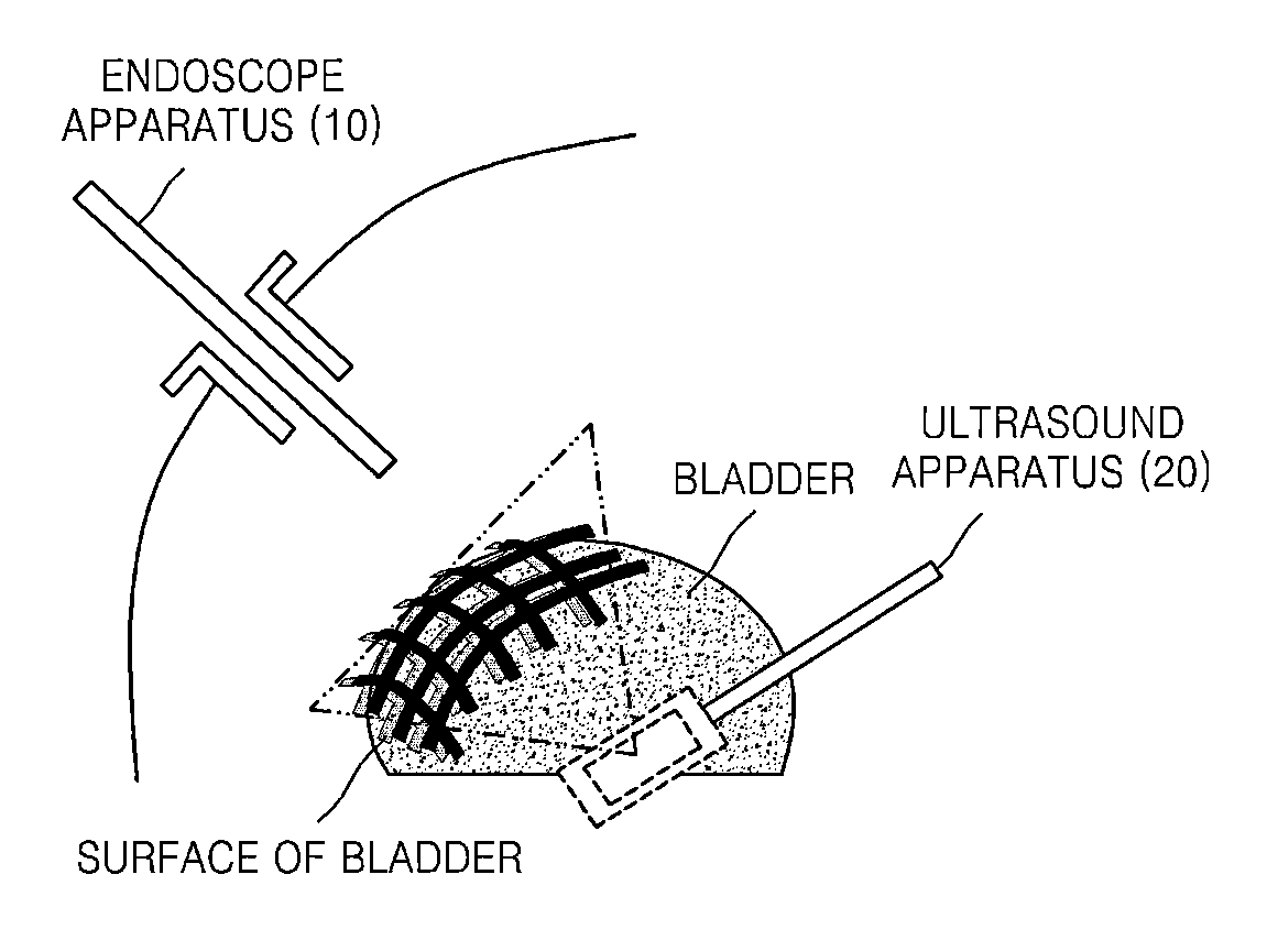
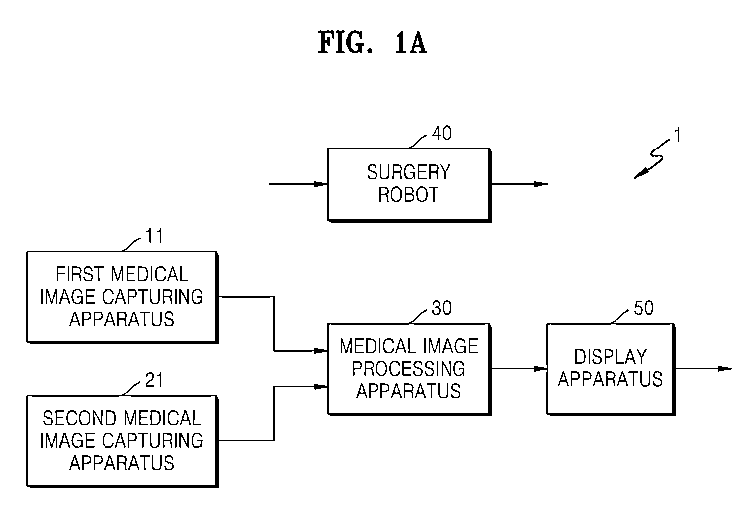
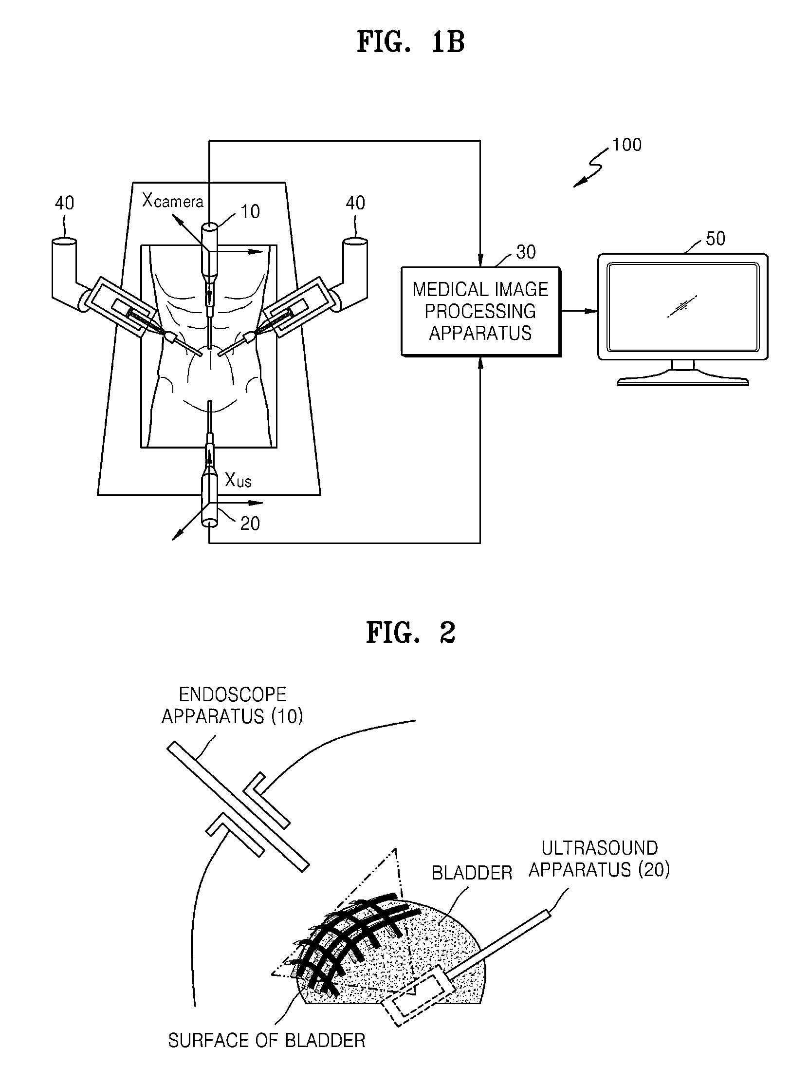
[0028]FIG. 1A is a block diagram of a robotic surgery system 1 according to an embodiment of the present disclosure. Referring to FIG. 1A, the robotic surgery system 1 may include, for example, a first medical image capturing apparatus 11, a second medical image capturing apparatus 21, a medical image processing apparatus 30, a surgery robot 40, and a display apparatus 50. In FIG. 1A and corresponding text, only hardware components associated with the current embodiment are described to keep aspects of the current embodim...


PUM


Abstract

A synthesis image in which medical images captured using different medical image capturing apparatuses are registered is generated by mapping the medical images to each other. The synthesis image may be used for image guidance while a diagnosis or a robotic surgery of a patient is being performed.

Description

CROSS-REFERENCE TO RELATED APPLICATIONS[0001]This application claims the priority benefit of Korean Patent Application No. 10-2011-0076993, filed on Aug. 2, 2011, in the Korean Intellectual Property Office, the disclosure of which is incorporated herein in its entirety by reference.BACKGROUND[0002]1. Field[0003]One or more embodiments of the present disclosure relate to a method and apparatus for processing a medical image, and a robotic surgery system using image guidance.[0004]2. Description of the Related Art[0005]Unlike manually performed abdominal operations, robotic surgeries allow surgeons to view a part to be operated on while inside the body of a patient through a screen display of a monitor instead of having to directly view the part to be operated on with the naked eye. While surgeons perform robotic surgeries after having perceived a part to be operated on using computed tomography (CT) images, magnetic resonance imaging (MRI) images, and ultrasound images, there is a li...

Claims


Application Information

Patent Timeline
Patent Type & Authority Applications(United States)
IPC IPC(8): A61B1/04G06K9/46A61B8/00A61B19/00A61B5/055A61B6/03
CPCA61B19/5212A61B2019/5227A61B2019/5289A61B2019/5297A61B19/2203A61B6/5247A61B6/506A61B6/5229A61B6/032A61B6/50A61B2019/5291A61B2090/368A61B2090/364A61B90/361A61B34/30A61B2090/371A61B2090/365A61B5/055A61B8/00G06T19/00
Inventor PARK, DONG-RYEOLKIM, YEON-HO
Owner SAMSUNG ELECTRONICS CO LTD
Why Eureka
Try Eureka