

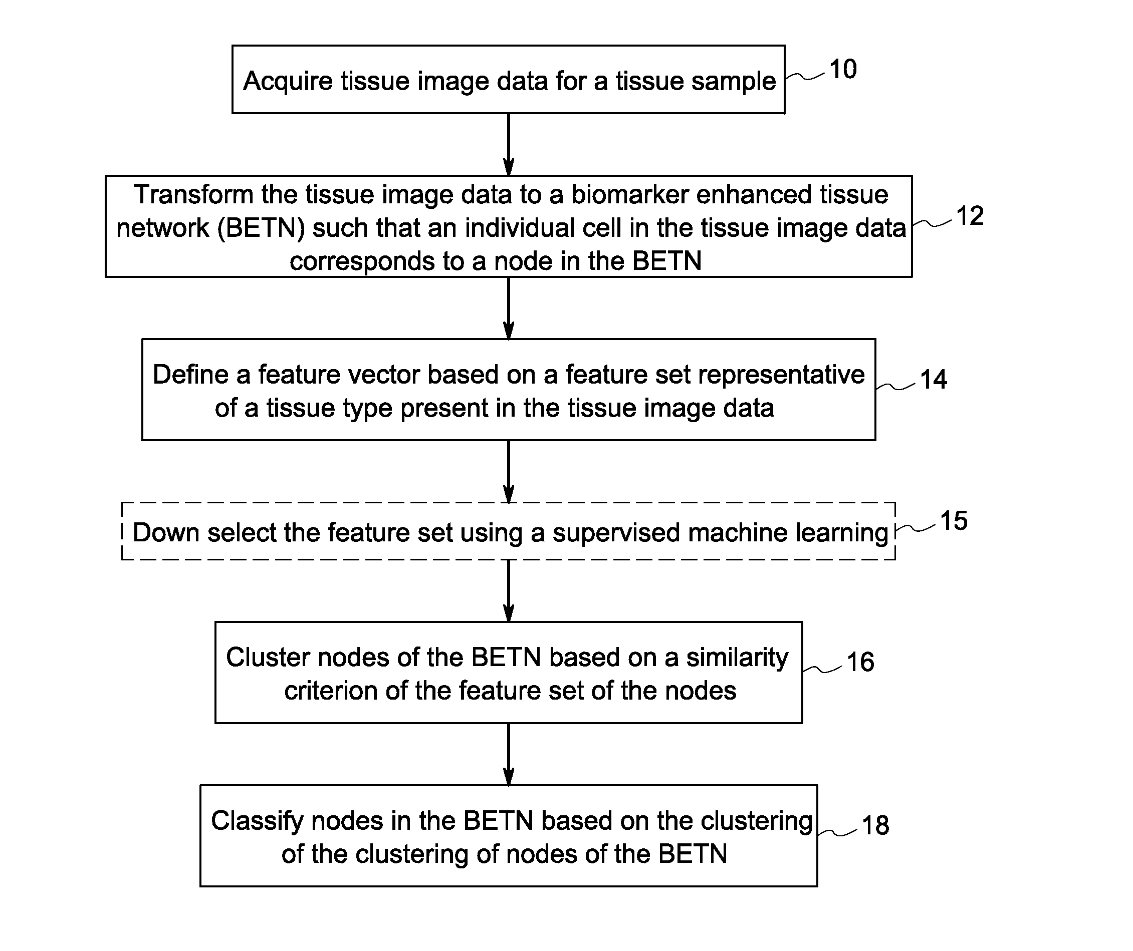
Systems and methods for tissue classification
a tissue classification and system technology, applied in the field of tissue images, can solve the problems of low reproducibility, high inefficiency and user variability of methods, and manual methods that cannot utilize the complex information that can be gained
- Summary
- Abstract
- Description
- Claims
- Application Information
AI Technical Summary
Benefits of technology
Problems solved by technology
Method used
Image

Examples
example
[0087]An algorithm was performed for feature selection. Using the 18 features listed below, with the addition of preliminary clustering features as genes, chromosomes of features were mutated, crossed over, and then selected using an SVM as a fitness function. Mutation and crossover was performed by taking two parent chromosomes (paired at random), keeping their common genes, then selecting a gene from the first parent only with a 40% chance, from the second parent only, another 40% chance, and a 20% chance of a random gene mutation (neither parent). Also, there was a small chance of dropping any gene at random, including a common one, to prevent premature convergence. The child chromosome size would be within a range created by the difference of the two parents' sizes added or subtracted to the parent with the higher fitness. The results shows that the clustering at threshold 40 and area of the cell tied with the most appearance in the final round, with area of the nucleus, mean cy...
PUM
 Abstract
Description
Claims
Application Information