Radiation imaging system, method for taking continuous radiographic image, and radiation image detecting device

a radiation imaging and continuous radiographic technology, applied in the field of radiation imaging, can solve the problems of affecting the accuracy of the detection of radiation, so as to achieve the effect of easy selection of dose detection sensors

Active Publication Date: 2013-03-28
FUJIFILM CORP
View PDF11 Cites 44 Cited by
  • Summary
  • Abstract
  • Description
  • Claims
  • Application Information

AI Technical Summary

Benefits of technology

[0028]According to the present invention, the radiation imaging system has the irradiation area map that defines the irradiation area of each small image capture field. Thus, it is possible to easily select the dose detection sensors belonging to the irradiation area in the continuous radiography.

Problems solved by technology

Therefore, it is difficult to optimize the X-ray dose in each of the division exposure positions.
Both the systems bring about increase in size and cost and a complicated and long-time process.
Thus, the AEC may be performed improperly.

Method used


Image

  • Radiation imaging system, method for taking continuous radiographic image, and radiation image detecting device
  • Radiation imaging system, method for taking continuous radiographic image, and radiation image detecting device
  • Radiation imaging system, method for taking continuous radiographic image, and radiation image detecting device

Examples


Embodiment Construction

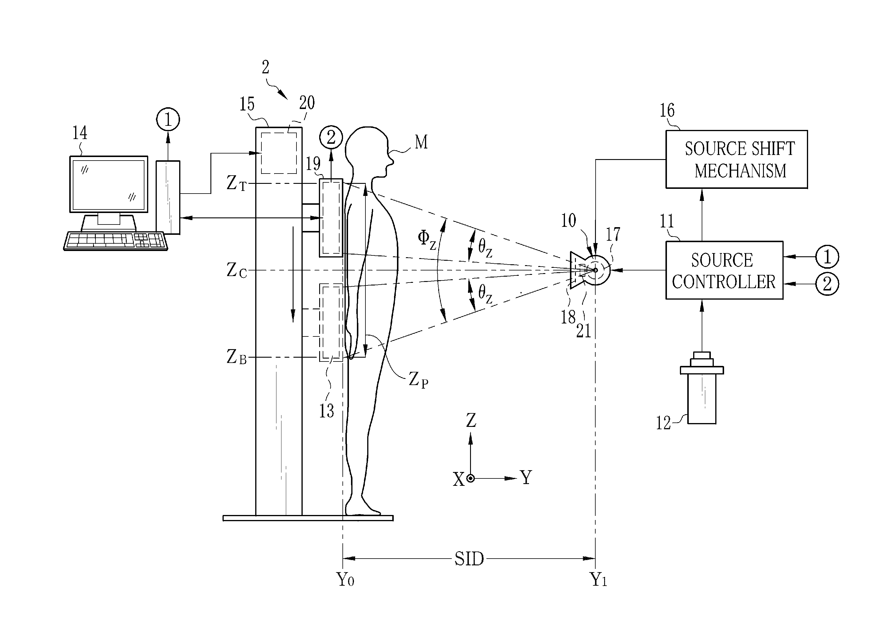
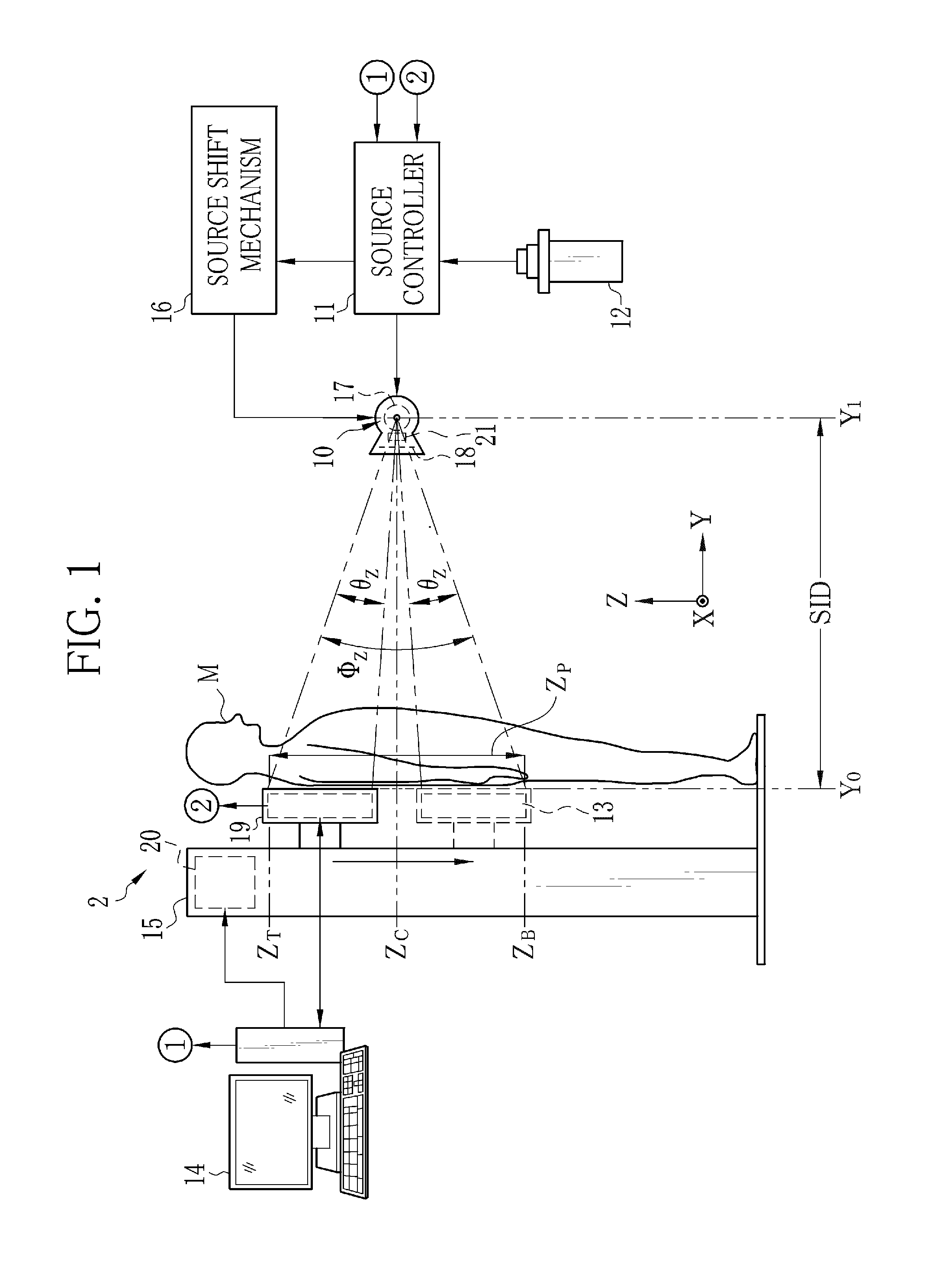
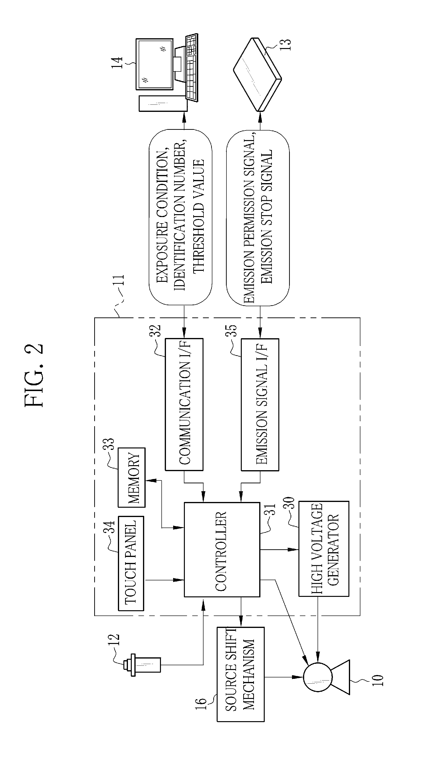
[0054]As shown in FIG. 1, an X-ray imaging system 2 is constituted of an X-ray source 10, a source controller 11, an exposure switch 12, an electronic cassette 13, a console 14, and an upright imaging support 15. The source controller 11 controls the operation of the X-ray source 10. The exposure switch 12 commands the start of X-ray emission. The electronic cassette 13, being a radiation image detecting device, detects X-rays having passed through a patient M to output an X-ray image. The console 14 controls the operation of the electronic cassette 13, and supplies various image processes to the X-ray image. The upright imaging support 15 is used in taking a radiograph of the patient M in a standing position. The X-ray source 10 is moved and set in a desired orientation and position by a source shift mechanism 16 or the like.

[0055]The X-ray source 10 has an X-ray tube 17 for emitting the X-rays, and a collimator 18 for limiting an irradiation field of the X-rays emitted from the X-...


PUM


Abstract

In continuous radiography, while a patient stands in front of an imaging support, a total image capture field is determined. The total image capture field is divided into small image capture fields. A map scaling section scales up or down a full spine irradiation area map in accordance with the size of the total image capture field. A map dividing section divides the scaled map into small maps corresponding to the small image capture fields. In each division exposure, a detection pixel selector selects one or more detection pixels belonging to an irradiation area defined by the small map, out of all detection pixels distributed in an imaging surface of an electronic cassette. If an integration value of a detection signal from the selected detection pixel reaches a threshold value, X-ray emission is stopped. Division X-ray images obtained by the division exposures are merged into a single continuous X-ray image.

Description

BACKGROUND OF THE INVENTION[0001]1. Field of the Invention[0002]The present invention relates to a radiation imaging system for taking a continuous radiographic image, a method for taking the continuous radiographic image, and a radiation image detecting device.[0003]2. Description Related to the Prior Art[0004]In a medical field, a radiation imaging system, for example, an X-ray imaging system using X-rays is widely known. The X-ray imaging system is constituted of an X-ray generation apparatus for generating the X-rays, and an X-ray image capturing apparatus for taking an X-ray image of a patient's body. The X-ray generation apparatus includes an X-ray source for emitting the X-rays to the body, a source controller for controlling the operation of the X-ray source, and an exposure switch for issuing an emission start signal of the X-rays. The X-ray image capturing apparatus includes an X-ray image detecting device for detecting the X-ray image based on the X-rays passed through th...

Claims


Application Information

Patent Timeline
Patent Type & Authority Applications(United States)
IPC IPC(8): A61B6/10
CPCA61B6/5241A61B6/542A61B6/548A61B6/4233A61B6/06A61B6/544
Inventor KAMIYA, TAKESHI
Owner FUJIFILM CORP
Why Eureka
Try Eureka