Automated Vertebral Body Image Segmentation for Medical Screening

a vertebral body and medical screening technology, applied in the field of computerized medical systems, can solve the problems of immobility, pain, mortality, costing billions of dollars annually, and low screening complian

Inactive Publication Date: 2013-03-28
RADNOSTICS
View PDF15 Cites 34 Cited by
  • Summary
  • Abstract
  • Description
  • Claims
  • Application Information

AI Technical Summary

Problems solved by technology

Osteoporosis is a condition of decreased bone mass that leads to fractures and negatively impacts millions of people, causing immobility, pain, and mortality, and costing billions of dollars annually.
Although an effective technique (DXA) for early diagnosis exists and the condition is treatable, screening compliance is low.
However, this is time consuming

Method used


Image

  • Automated Vertebral Body Image Segmentation for Medical Screening
  • Automated Vertebral Body Image Segmentation for Medical Screening
  • Automated Vertebral Body Image Segmentation for Medical Screening

Examples


Embodiment Construction

[0038]For better organization the description is broken down into subsections related to different portions of the system.

Vertebral Body Segmentation

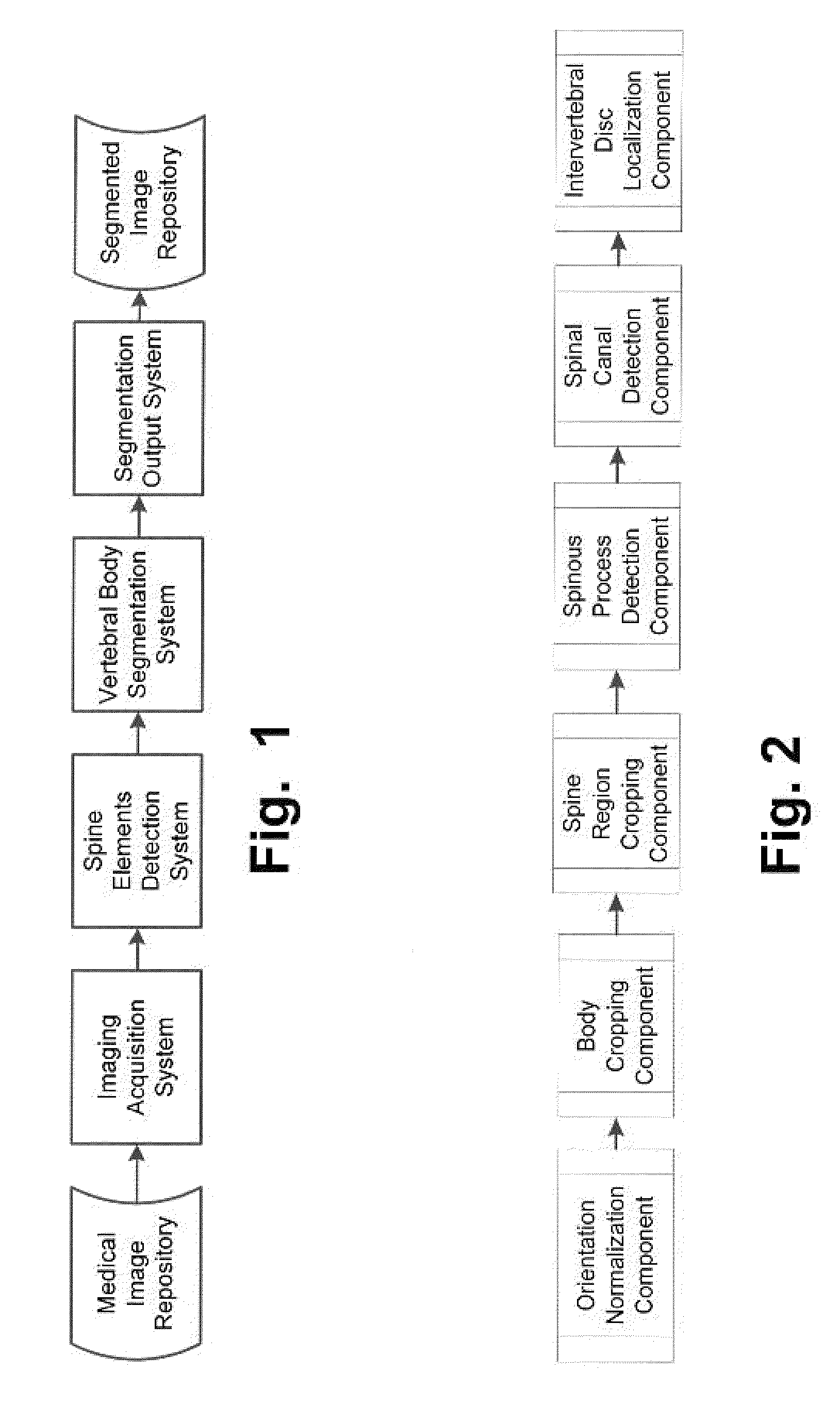
[0039]FIG. 1 is a block diagram illustrating the high, level workflow of the software pipeline developed to implement automated vertebral body segmentation. The imaging acquisition system retrieves the CT study from the medical image repository (e.g. the PACS system) and the spine elements detection system identifies the location of the spine elements in the study images. Next, the vertebral body segmentation system labels the cortex and the trabecular bone of each vertebral body. Finally, the segmentation output system stores the results in the segmented image repository (e,g, a hard drive).

Spine Element Detection System

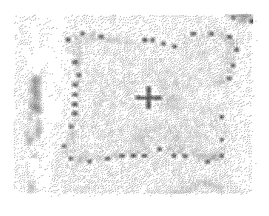
[0040]FIG. 2 is a flow chart illustrating the workflow of the spine elements detection system of FIG. 1, which isolates the spine region and detects the spinal canal and intervertebral discs in the CT study.
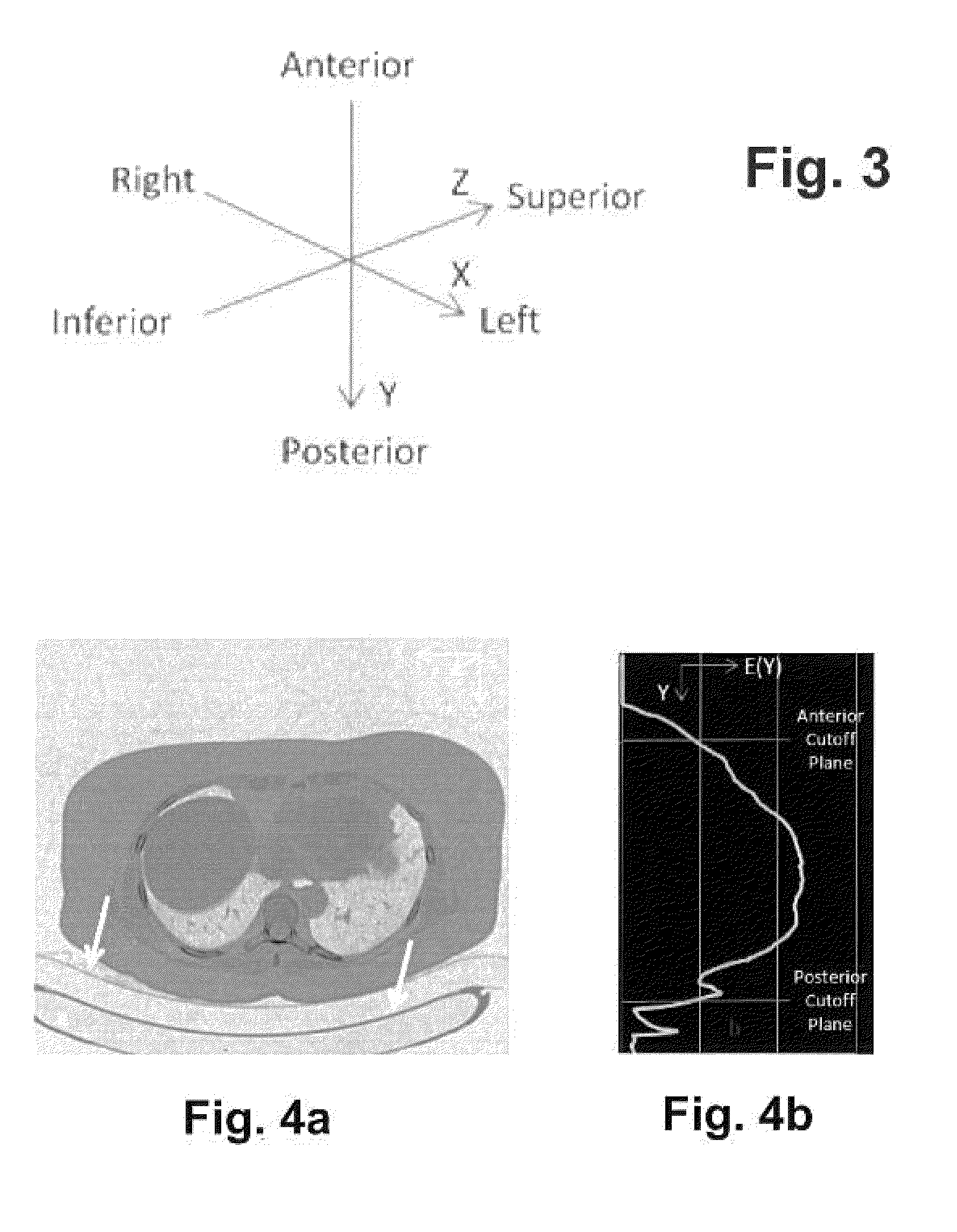
Orientation ...


PUM


Abstract

A method Is disclosed for fully automated segmentation of human vertebral body images in a CT (computerized tomography) study with no user interaction and no phantoms, which has resiliency to, anatomical abnormalities, and protocol and scanner variations. The method was developed to enable automated detection of osteoporosis in CT studies performed for other clinical reasons. Testing with 1.044 abdominal CTs from multiple sites, resulted in detection of 96.3% of the vertebral, bodies and 1% false positives. Of the detected vertebral bodies, 83.3% were segmented adequately for sagittal plane quantitative evaluation of vertebral, fractures indicative of osteoporosis. Improved results were observed when selecting the best sagittal plane of 3 for each vertebra, yielding a segmentation success rate of 85.4%. The method is preferably implemented in software as a building block in a system for automated osteoporosis detection.

Description

CROSS REFERENCES[0001]This application is a nonprovisional of U.S. Provisional Application Ser. No. 61 / 496,710, filed Jun. 14, 2011, entitled “AUTOMATED VERTEBRAL BODY SEGMENTATION,” the disclosure of which application is hereby incorporated by reference herein in its entirety.BACKGROUND OF THE INVENTION[0002]The present invention relates generally to computerized medical systems for diagnosing the human body and, more particularly, concerns procedures for analyzing images of a volume containing the spine to isolate the spine from its surroundings and to isolate the various components of the spine.[0003]Osteoporosis is a condition of decreased bone mass that leads to fractures and negatively impacts millions of people, causing immobility, pain, and mortality, and costing billions of dollars annually. Although an effective technique (DXA) for early diagnosis exists and the condition is treatable, screening compliance is low. Preliminary diagnosis of osteoporosis is achievable fay ins...

Claims


Application Information

Patent Timeline
IPC IPC(8): G06K9/34
CPCG06F19/321G06K9/34G16H30/40G16H30/20
Inventor BLUMFIELD, ANTHONYBLUMFIELD, EINAT
Owner RADNOSTICS
Why Eureka
Try Eureka