Radiation imaging apparatus and control method thereof, and radiation imaging system

a radiation imaging and control method technology, applied in the direction of radiation controlled devices, instruments, therapy, etc., can solve the problems of inability to obtain desirable image quality, inability of x-ray sensors to detect x-ray dose at an appropriate position in the object area, and inability to appropriately perform aec, etc., to achieve favorable image quality and simplify the structure of the radiation imaging apparatus

Active Publication Date: 2013-08-08
FUJIFILM CORP
View PDF5 Cites 39 Cited by
  • Summary
  • Abstract
  • Description
  • Claims
  • Application Information

AI Technical Summary

Benefits of technology

[0013]An object of the present invention is to provide a radiation imaging apparatus that has structure simpler than ever and can take a radiographic image of favorable image quality irrespective of a body portion to be imaged, a control method of the radiation imaging apparatus, and a radiation imaging system having the radiation imaging apparatus.
[0029]According to the present invention, exposure control is carried out based on the pixel value of the pixel in the image capturing field of the image detector. Thus, it is possible to simplify the structure of the radiation imaging apparatus. Furthermore, at least one typical low-value pixel is determined out of the pixels of the image capturing field, and the exposure control is performed based on the pixel value of the typical low-value pixel. Therefore, it is possible to obtain the radiographic image having favorable image quality, irrespective of the body portion to be image.

Problems solved by technology

Depending on the type, size, shape, or the like of a body portion, there may be cases where AEC cannot be performed appropriately, and desirable image quality cannot be obtained.
Thus, the X-ray sensors may not be able to detect the X-ray dose at an appropriate position in the object area, depending on the type, size, shape, or the like of the body portion.
Therefore, the apparatus of the Japanese Patent Laid-Open Publication No. 09-055298 may fail to perform AEC appropriately, depending on the type, size, shape, or the like of the body portion.
Thus, as with the Japanese Patent Laid-Open Publication No. 09-055298, the apparatus of the U.S. Pat. No. 7,433,445 may fail to perform appropriate AEC depending on the type, size, shape, or the like of the body portion, and fail to obtain favorable image quality.

Method used


Image

  • Radiation imaging apparatus and control method thereof, and radiation imaging system
  • Radiation imaging apparatus and control method thereof, and radiation imaging system
  • Radiation imaging apparatus and control method thereof, and radiation imaging system

Examples


Embodiment Construction

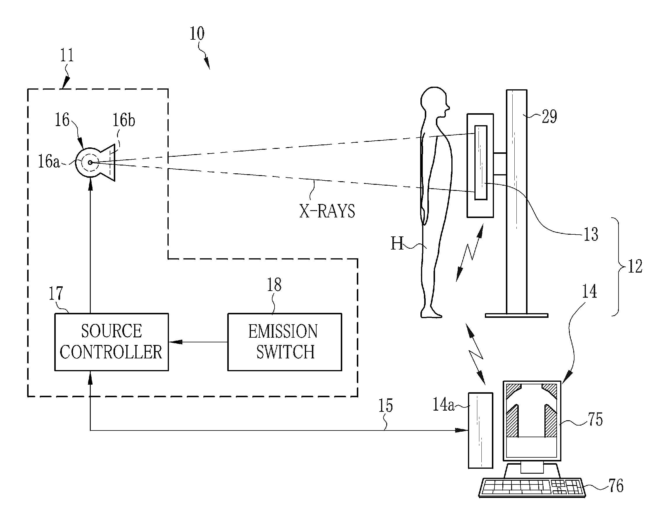
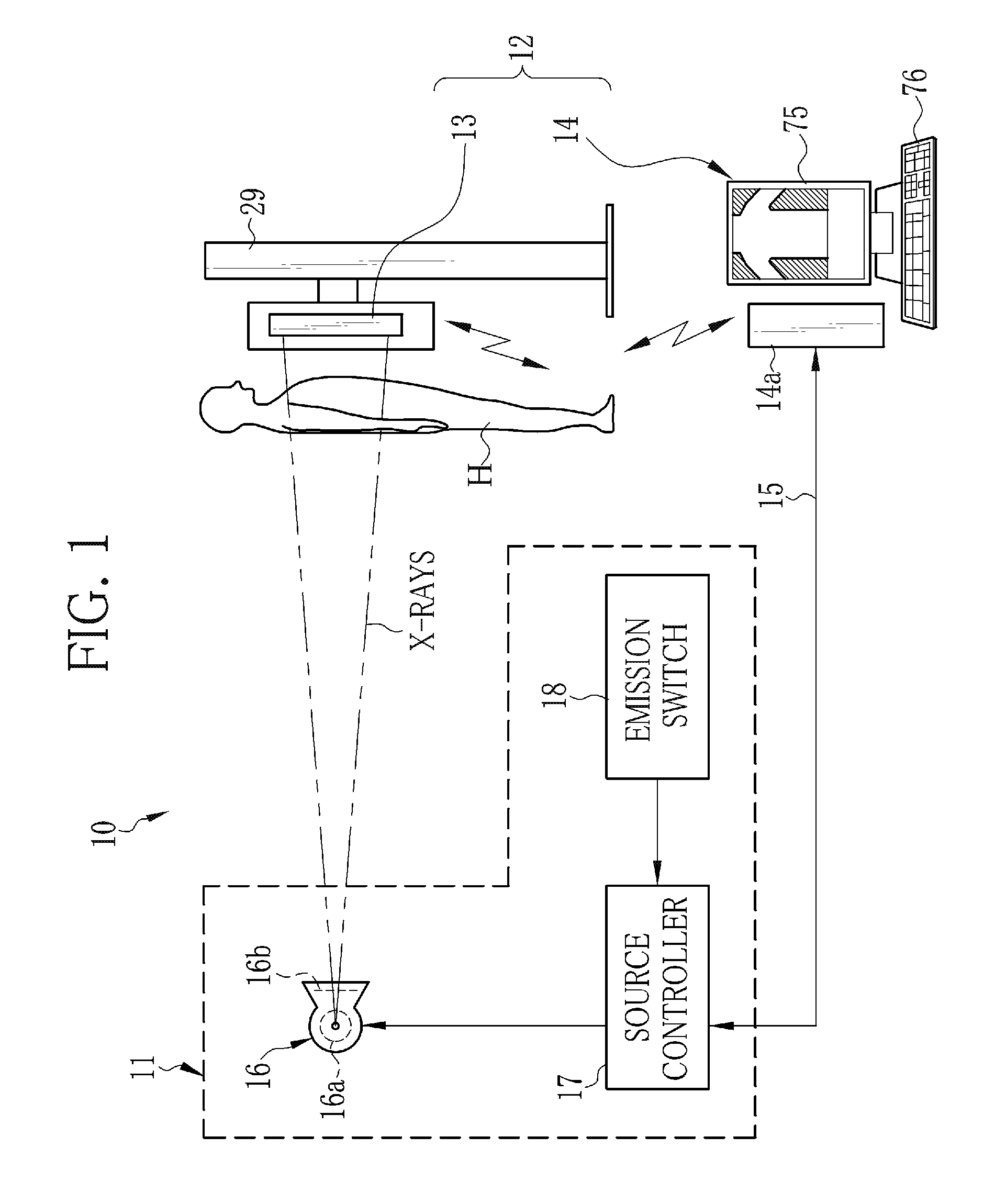
[0049]As shown in FIG. 1, an X-ray imaging system 10 is constituted of an X-ray generating apparatus 11 for generating X-rays and an X-ray imaging apparatus 12 for taking an X-ray image from the X-rays passed through a body portion (object) of a patient H. The X-ray imaging apparatus 12 includes an electronic cassette 13 for detecting the X-ray image and a console 14 that controls the electronic cassette 13 and performs image processing of the X-ray image. In the X-ray imaging system 10, the console 14 is communicatably connected to the X-ray generating apparatus 11 (concretely, a source controller 17) through a cable 15. The electronic cassette 13 and the console 14 are wirelessly communicatable with each other. The X-ray imaging system 10 carries out AEC (automatic exposure control) in which the console 14 stops X-ray emission from the X-ray generating apparatus 11 at the instant when an X-ray dose detected by the electronic cassette 13 has reached a predetermined value.

[0050]The ...


PUM


Abstract

An FPD detects an X-ray image of an object. The FPD includes a plurality of pixels arranged in its image capturing field. Each pixel receives X-rays emitted from an X-ray source, and outputs a pixel value in accordance with an X-ray dose applied thereto. A pixel determiner determines a minimum-value pixel out of the pixels based on the pixel values of the pixels. The minimum-value pixel is a pixel whose pixel value is the lowest. The pixel determiner sets the minimum-value pixel as an exposure control pixel. A comparator compares a first integrated value, which is an integrated value of the pixel values of the minimum-value pixel, with a predetermined first threshold value. The comparator performs X-ray emission control such that, when the first integrated value has reached the first threshold value, the X-ray source stops emitting the X-rays.

Description

BACKGROUND OF THE INVENTION[0001]1. Field of the Invention[0002]The present invention relates to a radiation imaging apparatus for taking a radiographic image from radiation passed through an object, a control method of the radiation imaging apparatus, and a radiation imaging system having the radiation imaging apparatus.[0003]2. Description Related to the Prior Art[0004]In a medical field, a radiation imaging system, for example, an X-ray imaging system using X-rays is widely known. The X-ray imaging system is constituted of an X-ray generating apparatus for applying the X-rays to an object (a body portion, for example, a chest of a patient), and an X-ray imaging apparatus for taking an X-ray image by reception of the X-rays passed through the object.[0005]In recent years, the X-ray imaging apparatus that uses a flat panel detector (FPD) as a detection panel, instead of an X-ray film or an imaging plate (IP), becomes widespread. The FPD has a matrix of pixels each of which produces...

Claims


Application Information

Patent Timeline
Patent Type & Authority Applications(United States)
IPC IPC(8): A61B6/00
CPCH05G1/38G01T1/026H05G1/56G01T7/005G01T1/02H05G1/44A61B6/4233G01T1/247A61B6/52A61B6/4283A61B6/542A61B6/548G01T1/1603H01L27/144H01L27/146H01L27/14605H01L27/14812A61B2560/0214A61B2560/0266A61N5/1071G01N2223/306
Inventor TSUJI, TETSUYA
Owner FUJIFILM CORP
Why Eureka
Try Eureka