Method and system to improve visceral adipose tissue estimate by measuring and correcting for subcutaneous adipose tissue composition

Active Publication Date: 2013-10-24
GENERAL ELECTRIC CO
View PDF3 Cites 0 Cited by
  • Summary
  • Abstract
  • Description
  • Claims
  • Application Information

AI Technical Summary

Benefits of technology

The patent describes a way to determine the amount of visceral adipose tissue (VAT) in a person's body using a dual-energy X-ray scan. This scan creates an image of the person and identifies different areas of interest on the image. These areas are then divided into smaller sections to create a soft tissue composition profile for each section. This profile identifies the edges of the person, the outer edge of their abdominal wall, the subcutaneous region, and the thickness of the subcutaneous layer. From this information, the amount of VAT can be calculated for each section. This method allows for more accurate measurement of VAT, which can help with the diagnosis of various medical conditions and the development of treatments for them.

Problems solved by technology

These devices often are not capable of providing accurate measurements of VAT because the actual fat content is not being measured, certain assumptions and / or estimates are made during the calculation process, and / or the devices are not exactly calibrated.
Also, reproducibility may be difficult, leading to inaccurate comparisons between examinations.
However, the use of these systems is often very costly and can expose a subject to high levels of ionizing radiation, for example, when using a CT imaging system.
Additionally, these imaging systems are not always available for clinical use and may have long scan times. Moreover, certain measurements are inaccurate in larger subjects.
However, these methods and systems often fail to accurately estimate SAT, thereby resulting in an inaccurate estimate of VAT.
It has been very difficult to determine the thickness of the subcutaneous fat region around the abdomen, especially near the buttocks, since the models used in the past do not take into account differences in the thickness of the subcutaneous fat region around the abdomen near the buttocks.

Method used


Image

  • Method and system to improve visceral adipose tissue estimate by measuring and correcting for subcutaneous adipose tissue composition
  • Method and system to improve visceral adipose tissue estimate by measuring and correcting for subcutaneous adipose tissue composition
  • Method and system to improve visceral adipose tissue estimate by measuring and correcting for subcutaneous adipose tissue composition

Examples


Embodiment Construction

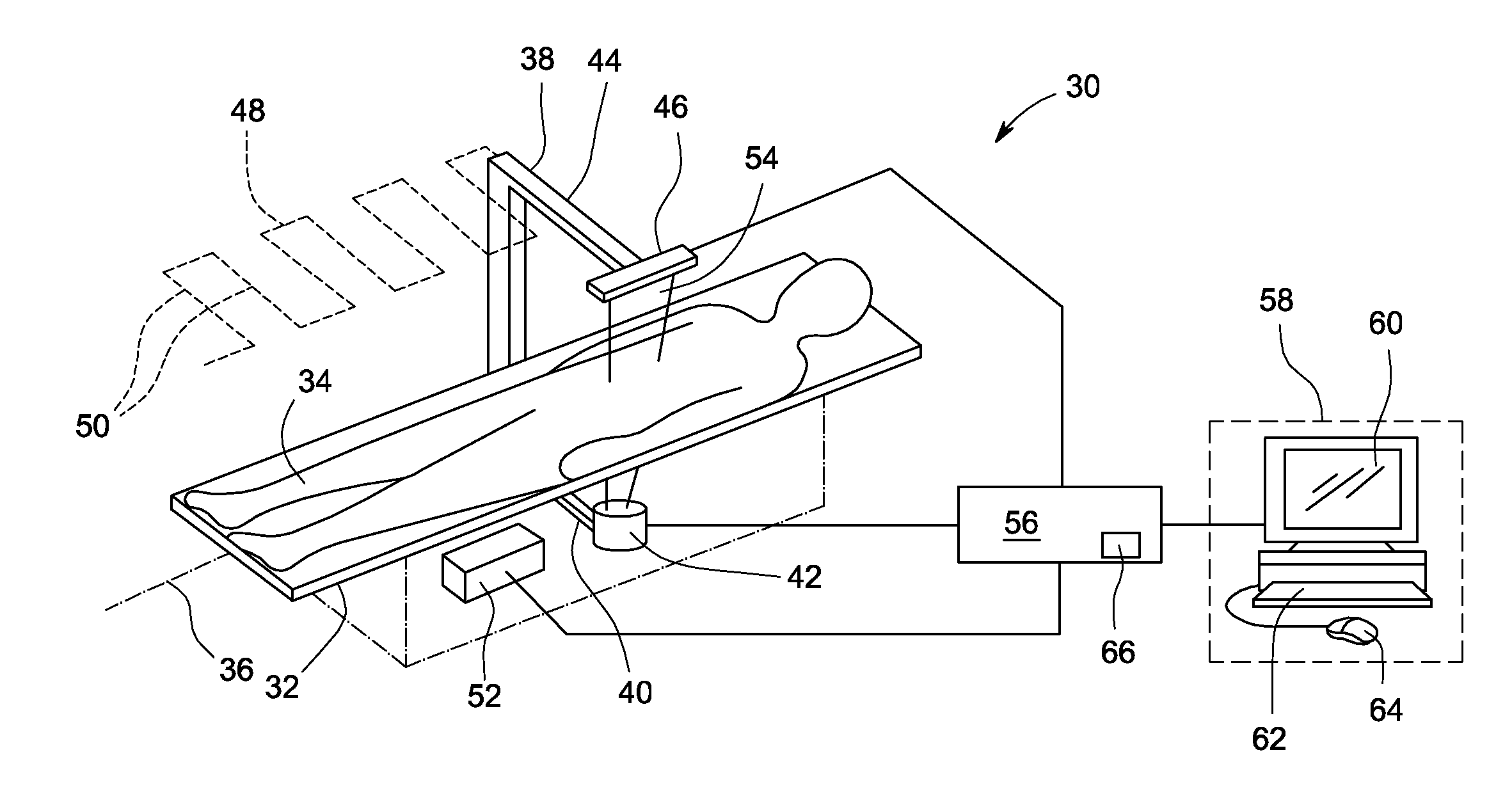
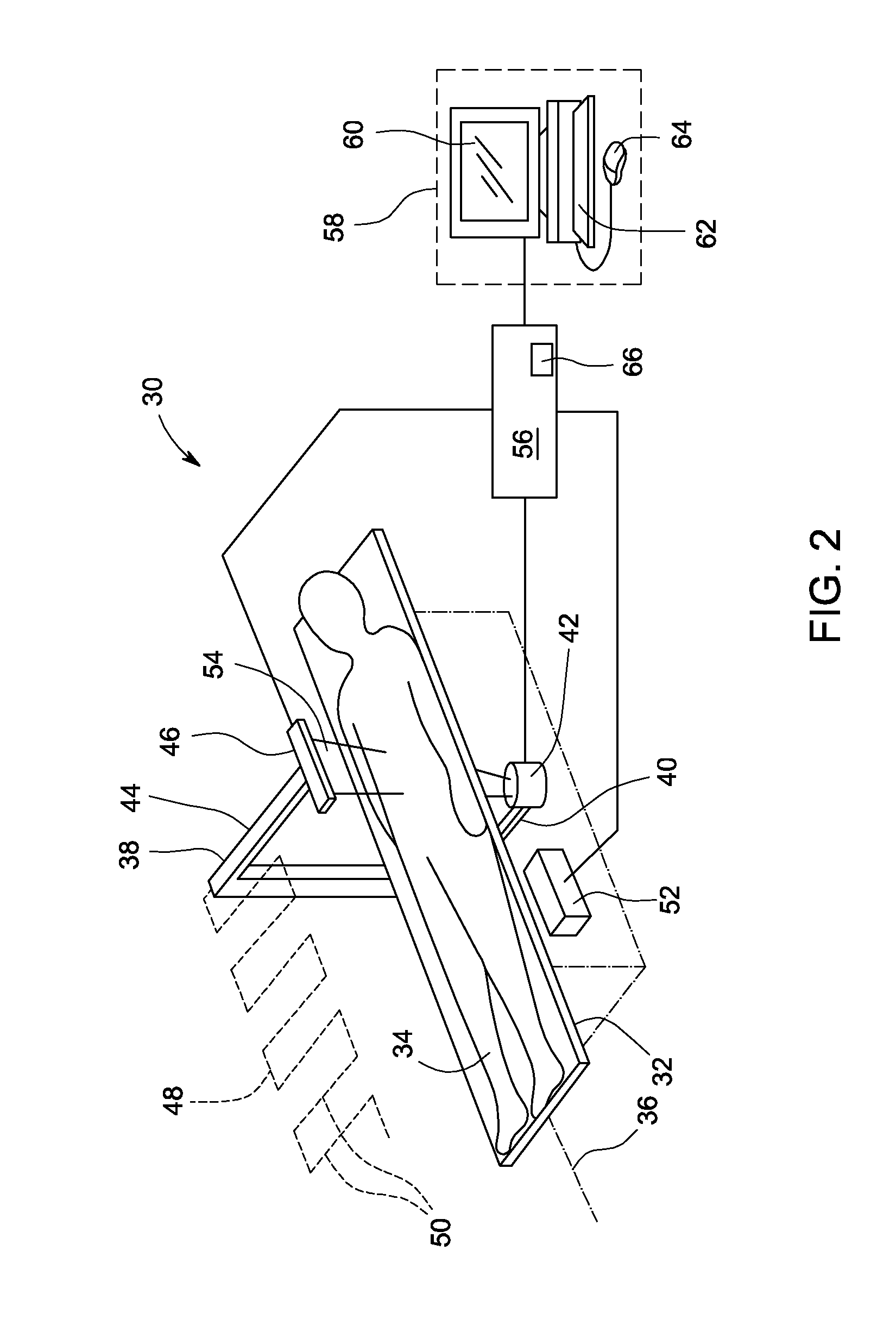
[0025]Exemplary embodiments of imaging methods and systems for scanning subjects to obtain tissue information, particularly soft tissue composition information and soft tissue thickness information, for providing and displaying visceral adipose tissue (VAT) information, are described in detail below. Different methods, systems, apparatus and models are used to measure the VAT of a subject in different regions of the subject's body.

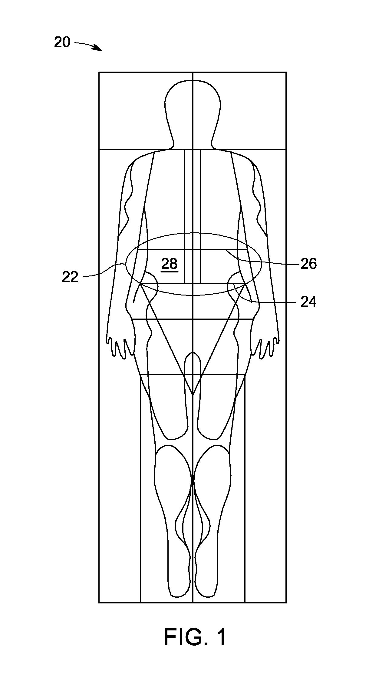
[0026]Referring now to the drawings, FIG. 1 illustrates a diagram of an exemplary embodiment of a dual-energy X-ray image 20 of a subject identifying a region of interest, such as an abdominal region of interest 22 to determine a VAT estimate of the subject. In particular, FIG. 1 is a full body dual-energy X-ray image 20 of a subject that may be generated from a scan of the entire body of the subject using a dual-energy X-ray absorptiometry (DXA) imaging system. The illustrated dual-energy X-ray image 20 is a dual-energy soft tissue image. The image 20 is ...


PUM


Abstract

A method and system for estimating the visceral adipose tissue (VAT) of a subject comprising performing a dual-energy X-ray scan of the subject; generating at least one image of the subject from the dual-energy X-ray scan; identifying a region of interest; dividing the region of interest into a plurality of sections; generating a soft tissue composition profile for each of the plurality of sections, wherein the soft tissue composition profile identifies an edge of the subject, an outer edge of an abdominal wall of the subject, a subcutaneous region and a subcutaneous thickness for each of the plurality of sections; determining a subcutaneous adipose tissue (SAT) composition for each of the plurality of sections, wherein a subcutaneous fat fraction is used to calculate the subcutaneous fat and lean masses and / or volumes; and determining a VAT based on the determined SAT composition for each of the plurality of sections.

Description

CROSS-REFERENCE TO RELATED APPLICATIONS[0001]This is a national stage application under 35 U.S.C. §371(c) of prior-filed, co-pending PCT patent application serial number PCT / US2011 / 068035, filed on Dec. 30, 2011, which claims priority to U.S. Provisional Patent Application No. 61 / 428,915, filed on Dec. 31, 2010, the contents of which are incorporated herein by reference in their entirety.BACKGROUND OF THE INVENTION[0002]This disclosure relates generally to imaging methods and systems, and more particularly to medical diagnostic imaging methods and systems that acquire and process tissue information for measuring the visceral adipose tissue (VAT) of a subject.[0003]Characteristics of a subject, such as body weight, fat mass, height, girth, gender, age, etc. are clinical descriptors useful by physicians to predict certain health risks that may increase or decrease mortality and morbidity risk. For example, the amount or type of abdominal fat, such as subcutaneous adipose tissue (SAT) ...

Claims


Application Information

Patent Timeline
IPC IPC(8): A61B5/00A61B6/03A61B5/055A61B6/00
CPCA61B5/4872A61B6/50A61B6/482A61B6/469A61B6/5217A61B6/032A61B5/055A61B6/4441A61B6/505A61B6/5235G16H50/30A61B6/5205
Inventor PAYNE, RANDALL KENNETH
Owner GENERAL ELECTRIC CO
Why Eureka
Try Eureka